Fundus color photo image blood vessel evaluation method and device, computer equipment and medium

A blood vessel and image technology, which is applied in the field of disease risk assessment in digital medical care, can solve problems such as inability to reflect the integrity of blood vessel segmentation results, and achieve good evaluation results

Active Publication Date: 2021-03-09
PING AN TECH (SHENZHEN) CO LTD
View PDF4 Cites 5 Cited by
  • Summary
  • Abstract
  • Description
  • Claims
  • Application Information

AI Technical Summary

Problems solved by technology

[0004] In order to overcome the above technical problems, especially when evaluating the accuracy of blood vessel segmentation in the prior art, the defect that the integrity of the topological structure of the blood vessel segmentation results cannot be reflected, the following technical solutions are proposed:

Method used


Image

  • Fundus color photo image blood vessel evaluation method and device, computer equipment and medium
  • Fundus color photo image blood vessel evaluation method and device, computer equipment and medium
  • Fundus color photo image blood vessel evaluation method and device, computer equipment and medium

Examples


Embodiment Construction

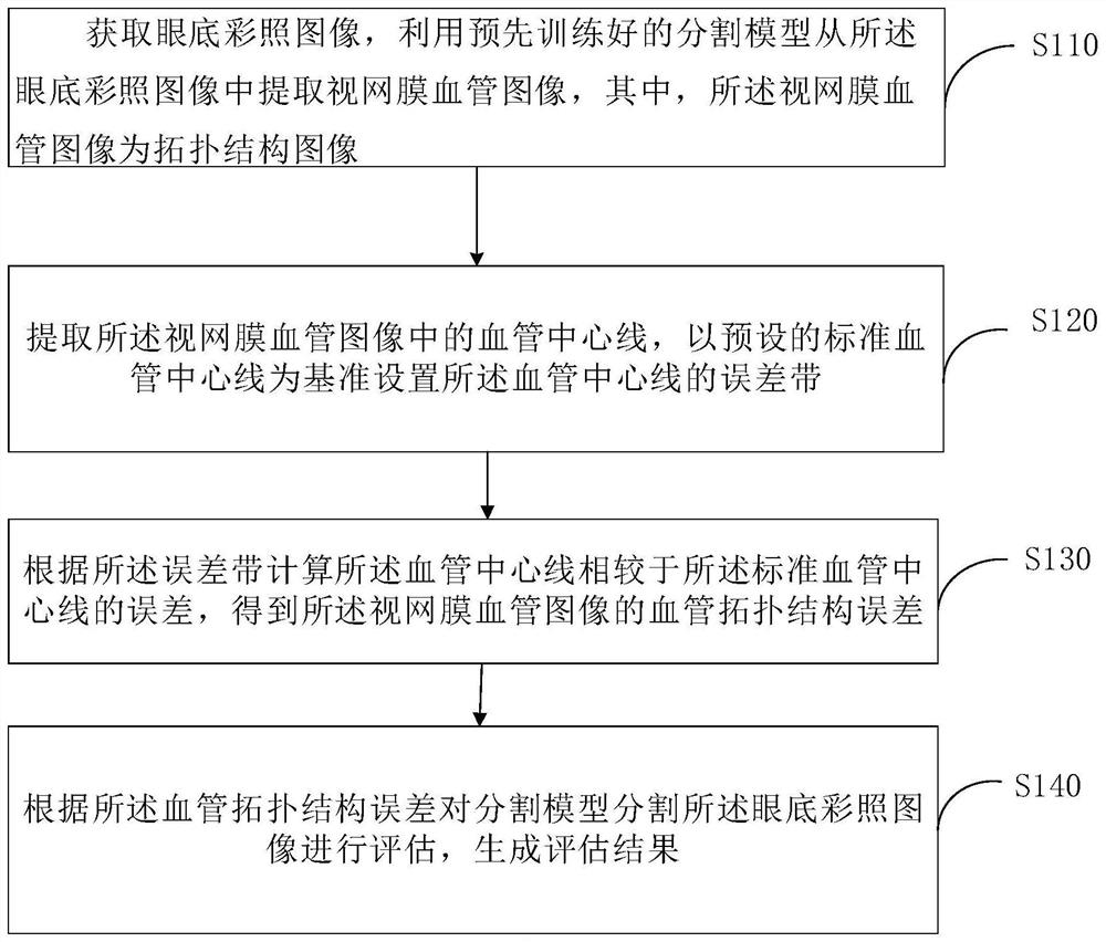
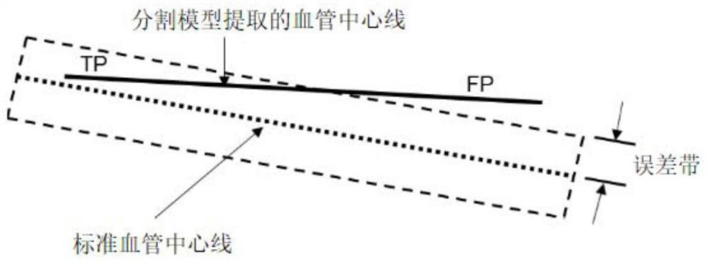
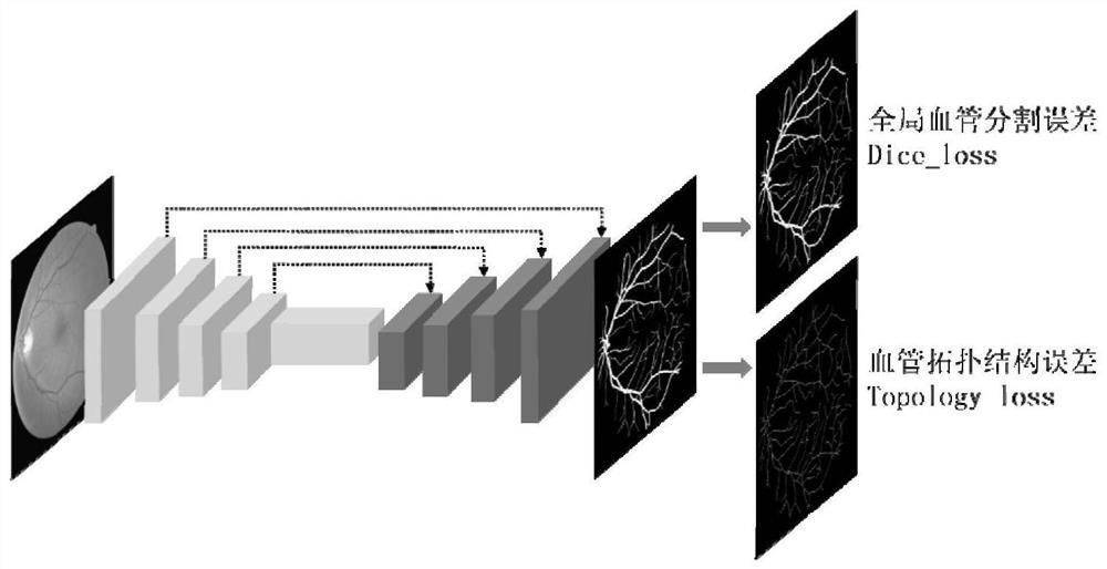
[0055]Embodiments of the present application are described in detail below, examples of which are shown in the drawings, wherein the same or similar reference numerals denote the same or similar elements or elements having the same or similar functions throughout. The embodiments described below by referring to the figures are exemplary only for explaining the present application, and are not construed as limiting the present application.

[0056] Those skilled in the art will understand that unless otherwise stated, the singular forms "a", "an", "said" and "the" used herein may also include plural forms. It should be further understood that the word "comprising" used in the specification of the present application refers to the presence of the features, integers, steps, operations, elements and / or components, but does not exclude the presence or addition of one or more other features, Integers, steps, operations, elements, components, and / or groups thereof. It will be unders...


PUM


Abstract

The invention relates to the technical field of disease risk assessment of digital medical treatment, and provides a fundus color photo image blood vessel evaluation method and device, computer equipment and a medium, and the method comprises the steps: extracting a retinal blood vessel image of a topological structure from a fundus color photo image through a pre-trained segmentation model; extracting a blood vessel center line in the retinal blood vessel image, setting an error band of the blood vessel center line by taking a preset standard blood vessel center line as a reference, and calculating an error of the blood vessel center line relative to the standard blood vessel center line according to the error band to obtain a blood vessel topological structure error of the retinal bloodvessel image; evaluating the fundus color photo image segmented by the segmentation model according to the vascular topological structure error, and generating an evaluation result, so as to evaluatethe integrity and accuracy of the blood vessel topological structure, and achieve a better evaluation effect.

Description

technical field [0001] The present application relates to the technical field of digital medical disease risk assessment, and in particular, the present application relates to a method, device, computer equipment and medium for assessing blood vessels in fundus color photograph images. Background technique [0002] Fundus color photography is a non-invasive, non-contact imaging technology that can directly observe retinal blood vessels, optic disc, macula and other fundus tissue structures, and is widely used in clinical screening and diagnosis of retinal fundus lesions. Among them, retinal blood vessels are the only vascular system that can be directly observed in the human body. Changes in retinal vessel morphology (such as vessel density, diameter, curvature, branch angle, etc.) are an important basis for early diagnosis and follow-up of many chronic cardiovascular diseases such as diabetes, hypertension, and kidney disease. [0003] At present, there are many studies on...

Claims


Application Information

Patent Timeline
Patent Type & Authority Applications(China)
IPC IPC(8): G06T7/00G06T7/11G06T7/155G06T7/62G06T7/68G06N3/08G06N3/04
CPCG06T7/0012G06T7/11G06T7/155G06T7/68G06T7/62G06N3/08G06T2207/30041G06T2207/30101G06N3/045
Inventor 柳杨吕彬吕传峰
Owner PING AN TECH (SHENZHEN) CO LTD
Why Eureka
Try Eureka