Method for establishing intracranial angiography enhanced three-dimensional stenosis analysis model

A contrast enhancement and three-dimensional model technology, applied in the field of image processing, can solve problems such as unfavorable positioning and analysis of intracranial vascular lesions, unfavorable intracranial blood vessels, and inability to obtain the analysis data of the degree of intracranial vascular stenosis intuitively and quickly. Achieve the effect of realizing 3D visualization and eliminating flow artifacts

Inactive Publication Date: 2021-03-16
XIAN CREATION KEJI CO LTD
View PDF0 Cites 1 Cited by
  • Summary
  • Abstract
  • Description
  • Claims
  • Application Information

AI Technical Summary

Problems solved by technology

However, two-dimensional images have limitations, which is not conducive to obtaining real information of intracranial blood vessels easily and quickly.
Moreover, from the images obtained by the above-mentioned imaging methods, it is impossible to intuitively and quickly obtain analysis data on the degree of intracranial vascular stenosis, which is not conducive to the clinical positioning and analysis of intracranial vascular lesion regions.

Method used


Image

  • Method for establishing intracranial angiography enhanced three-dimensional stenosis analysis model
  • Method for establishing intracranial angiography enhanced three-dimensional stenosis analysis model
  • Method for establishing intracranial angiography enhanced three-dimensional stenosis analysis model

Examples


Embodiment Construction

[0030] The present invention will be described in further detail below in conjunction with specific examples, but the embodiments of the present invention are not limited thereto.

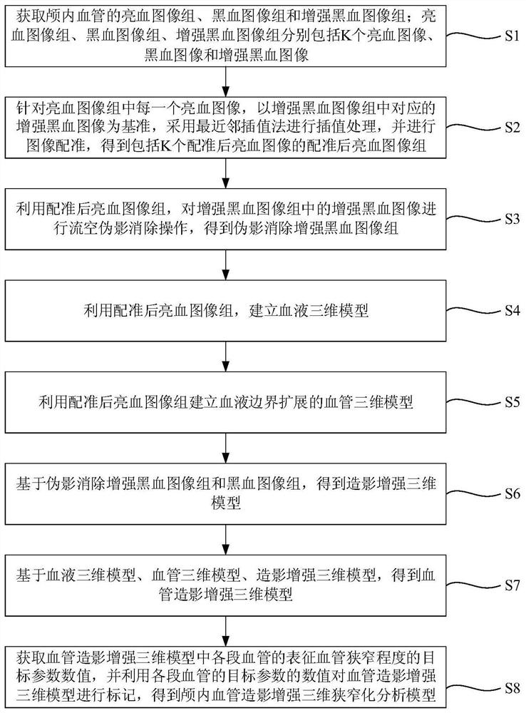
[0031] In order to obtain the real information of intracranial blood vessels and the analysis data about the degree of stenosis of intracranial blood vessels in a simple, fast and intuitive manner in clinical application, so as to analyze intracranial blood vessel lesions. An embodiment of the present invention provides a method for establishing a three-dimensional stenosis analysis model enhanced by intracranial angiography.

[0032] It should be noted that the execution subject of the method for establishing a three-dimensional stenosis analysis model enhanced by intracranial angiography provided in the embodiment of the present invention may be a device for establishing a three-dimensional stenosis analysis model enhanced by intracranial angiography. Can run in electronic equipment. Wherein, th...


PUM


Abstract

The invention discloses a method for establishing an intracranial angiography enhanced three-dimensional stenosis analysis model. The method comprises the following steps: acquiring a bright blood image group, a black blood image group and an enhanced black blood image group of intracranial vessels; carrying out interpolation processing on each bright blood image by utilizing a nearest neighbor interpolation method, and carrying out image registration to obtain a registered bright blood image group; carrying out flow empty artifact elimination operation on the enhanced black blood image by utilizing the registered bright blood image group to obtain an artifact-eliminated enhanced black blood image group; establishing a blood three-dimensional model and a blood vessel three-dimensional model by using the registered bright blood image group; obtaining a contrast enhanced three-dimensional model based on the artifact elimination enhanced black blood image group and the black blood image group; obtaining an angiography enhanced three-dimensional model; and obtaining values of target parameters representing the vascular stenosis degree of each section of blood vessel in the angiographyenhanced three-dimensional model, and marking the angiography enhanced three-dimensional model by utilizing the values of the target parameters of each section of blood vessel to obtain the intracranial angiography enhanced three-dimensional stenosis analysis model.

Description

technical field [0001] The invention belongs to the field of image processing, and in particular relates to a method for establishing an intracranial angiography-enhanced three-dimensional stenosis analysis model. Background technique [0002] According to statistics, the highest cause of human death is stroke, and stroke is caused by blockage or rupture of intracranial blood vessels, which affects the normal circulation of blood and causes infarction. Therefore, it is particularly important to evaluate the degree of intracranial vascular lesions and vascular stenosis. At present, methods based on lumen imaging are usually used to evaluate the degree of lesion and stenosis of intracranial vessels. Commonly used methods include digital subtraction angiography (Digital Subtraction Angiography, DSA), CT angiography (Computed Tomography Angiography, CTA ), magnetic resonance angiography (Magnetic Resonance Angiography, MRA) and high-resolution magnetic resonance angiography (Hi...

Claims


Application Information

Patent Timeline
Patent Type & Authority Applications(China)
IPC IPC(8): G06T11/00G06T7/00G06T7/13G06T7/136G06T7/33G06T3/40G06T5/30G06T17/00G16H30/20G16H30/40
CPCG06T3/4007G06T5/30G06T7/0012G06T11/008G06T17/00G06T2207/10088G06T2207/20016G06T2207/20024G06T2207/30101G06T2211/424G06T7/13G06T7/136G06T7/344G16H30/20G16H30/40
Inventor 贾思琪
Owner XIAN CREATION KEJI CO LTD
Why Eureka
Try Eureka