Supercharge Your Innovation With Domain-Expert AI Agents!

Device for separating neck vascular sheath from visceral sheath through body surface

A neck and blood vessel technology, applied in the field of medical equipment, can solve the problems of difficult operation, lack of blood circulation, soreness, etc., and achieve the effect of shortening operation time, expanding operation space, and easy operation.

Pending Publication Date: 2021-03-23
遵义医科大学第二附属医院
View PDF0 Cites 0 Cited by
  • Summary
  • Abstract
  • Description
  • Claims
  • Application Information

AI Technical Summary

Problems solved by technology

[0004] However, in the process of establishing the above-mentioned bone tunnel, using the two-finger method to establish a safe operating area must require the surgeon or assistant to maintain the operating area with at least one hand, which not only requires a small operating space, but also is more difficult to operate; Maintaining the operation area and maintaining the limbs will easily lead to blood stasis and soreness as the operation proceeds, resulting in a continuous decline in the stability of the finger maintenance, and it is difficult to control the risk of surgery. Currently, there is no public information about the auxiliary establishment of cervical vertebral bone tunnel

Method used


Image

  • Device for separating neck vascular sheath from visceral sheath through body surface
  • Device for separating neck vascular sheath from visceral sheath through body surface
  • Device for separating neck vascular sheath from visceral sheath through body surface

Examples


Embodiment

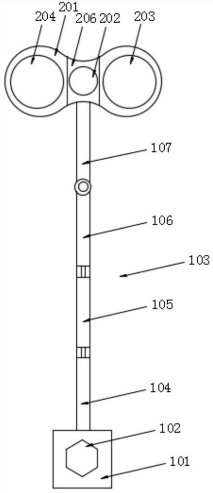
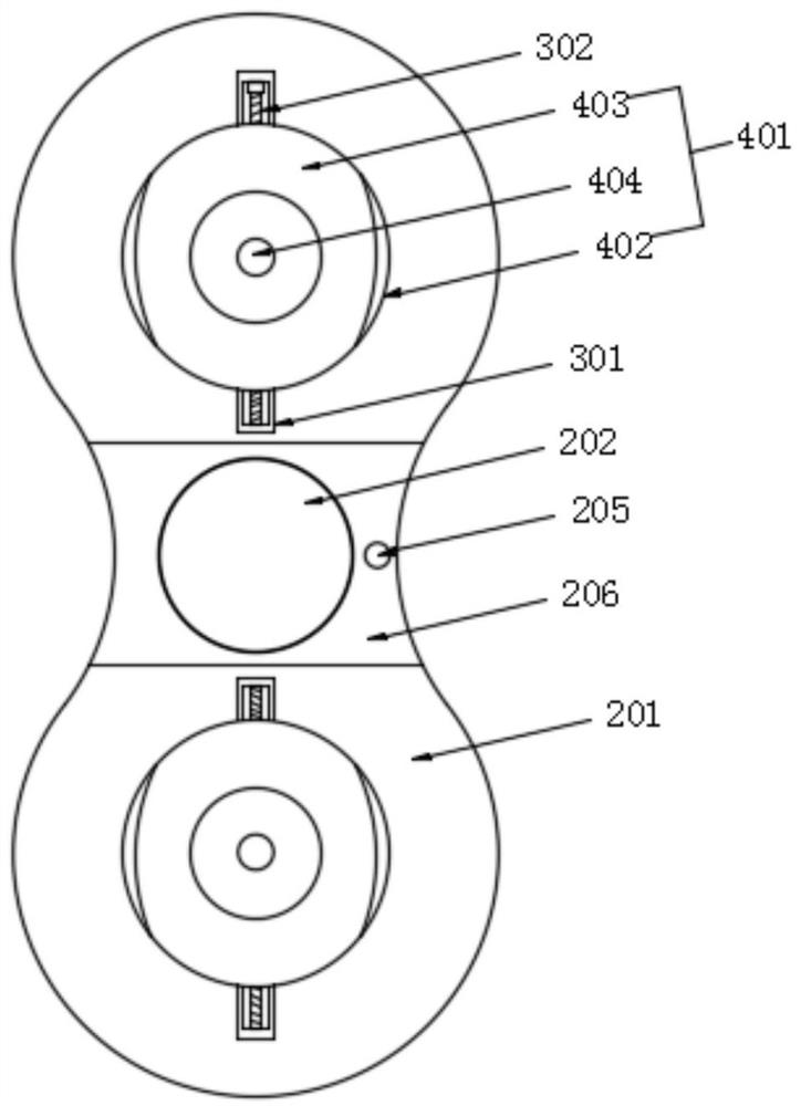
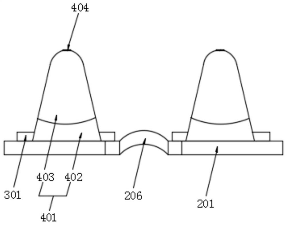
[0044] Example: a device for separating the cervical vascular sheath and visceral sheath through the body surface, such as figure 1 , figure 2 and Figure 7 As shown, it includes a fixed seat 101 , a mechanical arm 103 , a carrying platform 201 , an image processor 108 , and a controller 109 . The fixed seat 101 is connected to the carrying platform 201 through the mechanical arm 103 . The bottom surface of the bearing platform 201 is symmetrically provided with two positioning cones 401 distributed at intervals. The end of the positioning cone 401 away from the bearing platform 201 is provided with an ultrasonic probe 404. The output ends of the two ultrasonic probes 404 are connected to the input of the image processor 108. The output terminal of the image processor 108 is connected to the input terminal of the controller 109, and the output terminal of the controller 109 is connected to the display device. The carrying platform 201 is pierced with an operation port 202 a...


PUM


Abstract

The invention discloses a device for separating a neck vascular sheath from a visceral sheath through a body surface, and relates to the technical field of medical equipment. The device is characterized by comprising a fixed seat, a mechanical arm, a bearing table, an image processor and a controller, wherein the fixed seat is connected with the bearing table through the mechanical arm, the fixedseat is in screw-thread fit with a fixed bolt, two positioning cones distributed at intervals are symmetrically arranged on the bottom surface of the bearing table, ultrasonic probes are arranged at the ends, away from the bearing table, of the positioning cones, the output ends of the two ultrasonic probes are connected with the input end of the image processor, the output end of the image processor is connected with the input end of the controller, and the output end of the controller is connected with a display device; and an operation opening aligned with the intersection area of the two ultrasonic probes for ultrasonic scanning is formed in the bearing table in a penetrating mode. The device is simple and convenient in overall operation, does not need auxiliary personnel to maintain the operation area for a long time, expands the operation space, and can effectively control the operation risk by shortening the operation time and enhancing the stability of the operation environment.

Description

technical field [0001] The invention relates to the technical field of medical equipment, and more specifically, relates to a device for separating a cervical vascular sheath and a visceral sheath through a body surface. Background technique [0002] Cervical disc herniation is a common disease. The main reason is that on the basis of the pathology of intervertebral disc degeneration, the outer fibrous annulus of the intervertebral disc is further ruptured, and the nucleus pulposus tissue protrudes (or protrudes) and compresses the spinal nerve root or spinal cord, resulting in neck pain. , Numbness and weakness of limbs or even clinical symptoms of paralysis. [0003] Endoscopic anterior transvertebral bone tunnel technology is a new method for the treatment of cervical disc herniation. How to establish a bone tunnel directly to the posterior vertebral body through percutaneous minimally invasive surgery is an important step in this technology. During the operation, use th...

Claims


Application Information

Patent Timeline
IPC IPC(8): A61B17/00
CPCA61B17/00234A61B2017/00261
Inventor 廖文波杜迁孔维军曹广如辛志军张敏波
Owner 遵义医科大学第二附属医院
Features
  • R&D
  • Intellectual Property
  • Life Sciences
  • Materials
  • Tech Scout
  • Unparalleled Data Quality
  • Higher Quality Content
  • 60% Fewer Hallucinations
Learn More