Automatic mouse spermatogenic tube staging system based on tissue morphological analysis

A technology of tissue morphology and spermatogenesis, applied in image analysis, image data processing, instruments, etc., can solve problems such as difficult staging, achieve good classification accuracy and assist staging identification.

Pending Publication Date: 2021-03-23
NANJING UNIV OF INFORMATION SCI & TECH
View PDF2 Cites 0 Cited by
  • Summary
  • Abstract
  • Description
  • Claims
  • Application Information

AI Technical Summary

Problems solved by technology

Due to the large number of germ cell types and complex structures in the seminiferous duct, and the small difference between two consecutive spermatogenic phases, it is difficult to perform manual staging

Method used


Image

  • Automatic mouse spermatogenic tube staging system based on tissue morphological analysis
  • Automatic mouse spermatogenic tube staging system based on tissue morphological analysis
  • Automatic mouse spermatogenic tube staging system based on tissue morphological analysis

Examples


specific Embodiment

[0033] DETAILED DESCRIPTION Figure 4 The workflow of the mouse sequester automatic installment system is as follows:

[0034] 1. First, first, a mouse testicular slice is scanned by a mouse testicular slice, and the length and width is reduced by 20 times. The pre-segmentation result of the semacchaiosis; the division result is mapped to the original map using the bilayer interpolation method.

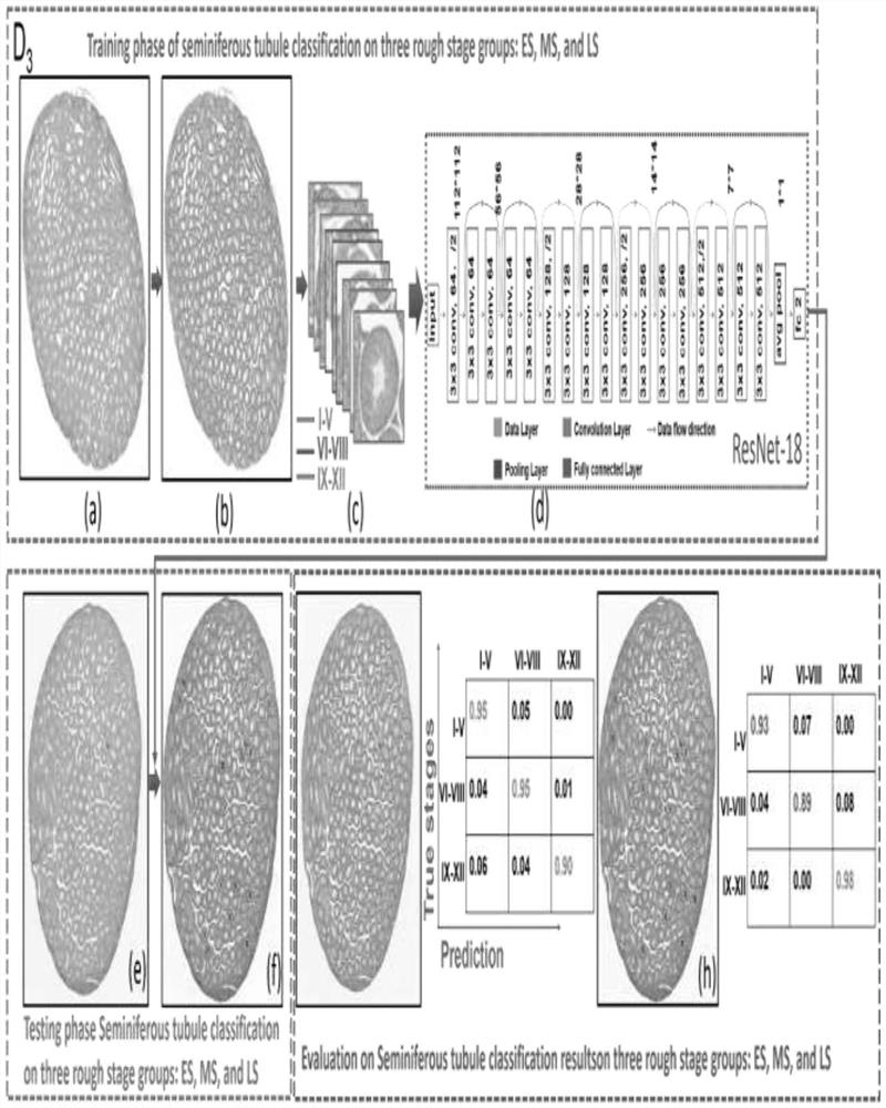
[0035] 2, then, the spermatin predishes predishes in the full scan image is subjected to classification in the depth convolutional neural network, resulting in the classification result of the I-VI, VII-VIII, ⅺ-ⅻ-ⅻ period, Such as figure 2 Said;

[0036] 3, once again, extract the spermatin classified from VII-VIII period, and use the depth convolutional neural network to the nucleus, and use the depth full consolidation neural network (UNET) to segment it, such as Figure 5 Said;

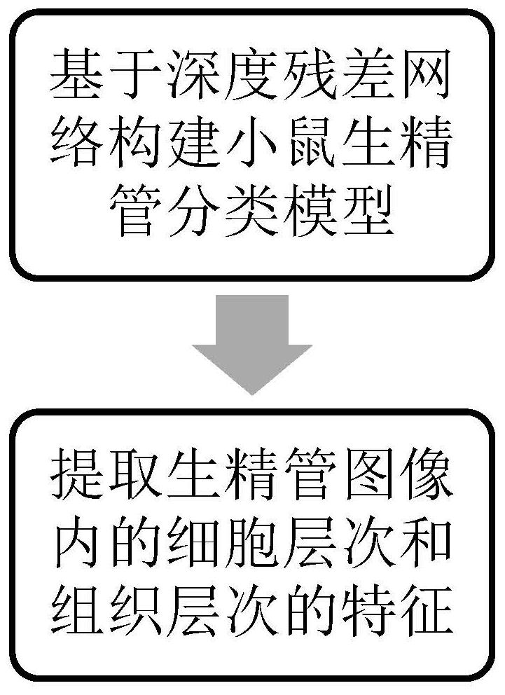
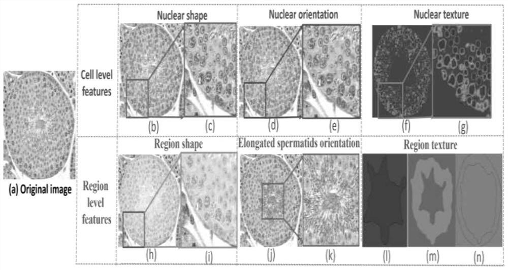
[0037] 4, finally, the characteristics of the cell level and tissue level are extracted for the organizatio...


PUM


Abstract

The invention discloses an automatic mouse spermatogenic tube staging system based on tissue morphological analysis. The method belongs to the field of machine learning and image processing. The method comprises the following specific steps: constructing a mouse spermatogenic tube classification model based on a deep residual network (ResNet); and extracting characteristics of a cell level and a tissue level in the spermatogenic tube image. According to the method, a deep learning method is used for automatically segmenting multiple types of spermatogenic tubes, multiple types of cells in thespermatogenic tubes and multiple types of regions in the spermatogenic tubes in the testis of the mouse; in addition, in the work of feature extraction and feature selection, long sperm direction features are manually designed on the basis of existing pathological features; finally, the classification accuracy of the selected features is good and conforms to the staging experience of pathologists,mutual interpretation can be effectively achieved, and the correctness of the selected features is fully verified; in addition, the successful establishment of the VI, VII-mVIII and lateVIII stagingsystems can provide quantitative information for pathologists during staging diagnosis, and assist the pathologists in staging identification.

Description

Technical field [0001] The present invention relates to the field of machine learning and image processing, and more particularly to mice based on tissue morphological analysis automatic installment systems. Background technique [0002] Since the testicular pathological structure of mammals is very similar, the early lesions of infertility caused by pathological defects in human reproductive are usually experimental in mouse testis. During sperm occurrence, a particular cell combination formed in a genital cell in different developments is referred to as a biological phase phase. By observing the characteristics of the cyclical phase-in-chore-in-chimetricular phase in the sperm occurrence process, the mice were divided into I-XII. Due to many reproductive cell types in the biostile, the structure is complex, and the two consecutive bins are different, which leads to a staging in artificial way. However, accurately dividing the sperm occurrence process, static complex dynamics of...

Claims


Application Information

Patent Timeline
Patent Type & Authority Applications(China)
IPC IPC(8): G06T7/00G06T7/11G06T7/49G06K9/62G06K9/46
CPCG06T7/0016G06T7/11G06T7/49G06V10/44G06F18/2135G06F18/2414G06F18/2411
Inventor 徐军鲁浩达
Owner NANJING UNIV OF INFORMATION SCI & TECH
Why Eureka
Try Eureka