Method and equipment for ultrasonography and image storage method for ultrasonography

A technology of ultrasound image and contrast-enhanced ultrasound, which is applied in the fields of ultrasound/sonic/infrasonic image/data processing, ultrasound/sonic/infrasonic diagnosis, ultrasound/sonic/infrasonic Permian technology, etc. It can reduce the complexity and difficulty of operations, reduce storage requirements, and improve work efficiency.

Pending Publication Date: 2021-03-26
SHENZHEN MINDRAY BIO MEDICAL ELECTRONICS CO LTD +1
View PDF6 Cites 1 Cited by
  • Summary
  • Abstract
  • Description
  • Claims
  • Application Information

AI Technical Summary

Problems solved by technology

[0006] In addition, under the current workflow framework, after the doctor opens the stored angiographic dynamic image, selects and saves the desired static image or dynamic video, he cannot browse the selected images or video on the same screen
You can only watch or browse the short films one by one, which is not conducive to intuitive comparison and analysis

Method used

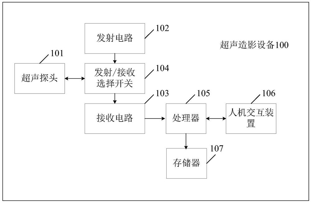
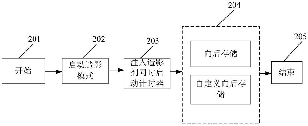
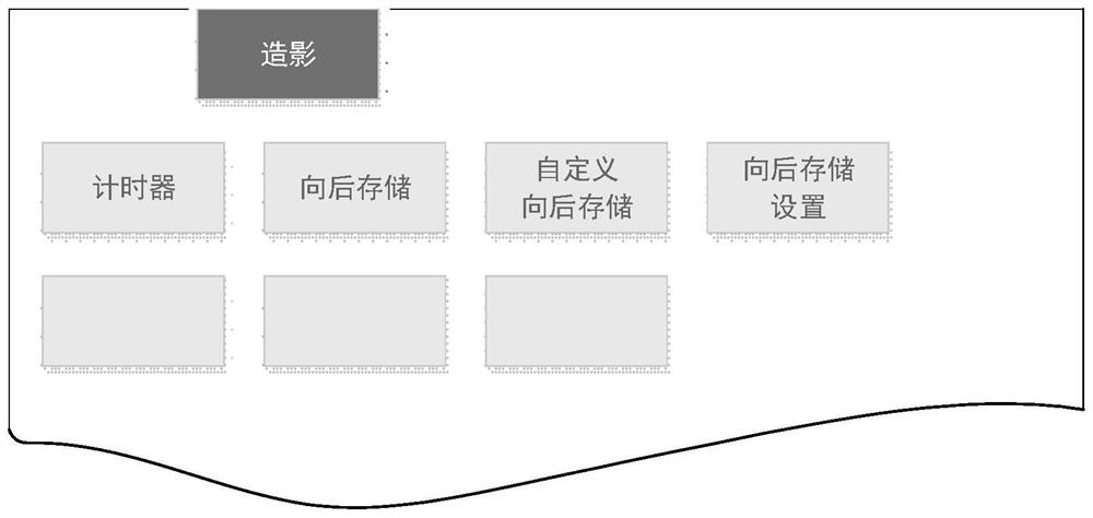

Image

  • Method and equipment for ultrasonography and image storage method for ultrasonography
  • Method and equipment for ultrasonography and image storage method for ultrasonography
  • Method and equipment for ultrasonography and image storage method for ultrasonography

Examples


Embodiment Construction

[0047] In order to make the objects, technical solutions and advantages of the present invention more apparent, exemplary embodiments according to the present invention will be described in detail below with reference to the accompanying drawings. Apparently, the described embodiments are only some embodiments of the present invention, not all embodiments of the present invention, and it should be understood that the present invention is not limited by the exemplary embodiments described here. Based on the embodiments of the present invention described in the present invention, all other embodiments obtained by those skilled in the art without creative efforts shall fall within the protection scope of the present invention.

[0048]In the following description, numerous specific details are given in order to provide a more thorough understanding of the present invention. It will be apparent, however, to one skilled in the art that the present invention may be practiced without...


PUM


Abstract

The invention provides a method for ultrasonography, an image storage method for ultrasonography and ultrasonography equipment. The method for ultrasonography comprises the steps: transmitting first ultrasonic waves to inspected tissue, so as to determine a target area; transmitting second ultrasonic waves to the contrast-microbubble-containing target area when the target area contains contrast microbubbles, so as to obtain an ultrasonic image at least comprising contrast images; receiving a first instruction for storing dynamic images of the ultrasonic image; and storing the dynamic images inresponse to the first instruction. The method further comprises the steps: when the dynamic images are stored, receiving a second instruction for storing one-frame or multi-frame static images and/ordynamic short films in the dynamic images; and storing the one-frame or multi-frame static images and/or dynamic short films in response to the second instruction. According to the methods and the equipment, the complexity and difficulty of inspection operations of doctors can be lowered, and the working efficiency is increased.

Description

technical field [0001] The present invention generally relates to the technical field of contrast-enhanced ultrasound, and more specifically relates to a method for contrast-enhanced ultrasound imaging, a method for storing images of contrast-enhanced ultrasound, and contrast-enhanced ultrasound equipment. Background technique [0002] Hepatic contrast-enhanced ultrasonography was the earliest and most widely used clinically, and the effect is also the most significant. This is closely related to the special blood supply mode of the liver which is different from other organs. The blood supply of the hepatic artery and the portal vein and the solid background of the liver make the liver the best target organ for contrast enhancement. Contrast-enhanced ultrasonography of the liver is divided into arterial phase, portal phase, and delayed phase, and the differential diagnosis is made according to the contrast characteristics of the lesion. In addition to helping to identify t...

Claims


Application Information

Patent Timeline
Patent Type & Authority Applications(China)
IPC IPC(8): A61B8/08A61B8/00
CPCA61B8/481A61B8/467A61B8/463A61B8/465
Inventor 桑茂栋章希睿王丰陈玉鑫刘学东
Owner SHENZHEN MINDRAY BIO MEDICAL ELECTRONICS CO LTD
Why Eureka
Try Eureka