Hip joint segmentation model building method using small sample image training and application thereof

A technology of segmentation model and establishment method, applied in the field of medical image processing, can solve problems such as poor segmentation results, and achieve the effect of accelerating network convergence and ensuring training effect.

Pending Publication Date: 2021-04-09
HUAZHONG UNIV OF SCI & TECH
View PDF0 Cites 6 Cited by
  • Summary
  • Abstract
  • Description
  • Claims
  • Application Information

AI Technical Summary

Problems solved by technology

[0005] Aiming at the defects and improvement needs of the prior art, the present invention provides a method for establishing a hip joint segmentation model using small sample image training and its application. The purpose is to effectively solve the problem that the existing hip joint segmentation method relies on Labeling data, technical problems of poor segmentation results

Method used


Image

  • Hip joint segmentation model building method using small sample image training and application thereof
  • Hip joint segmentation model building method using small sample image training and application thereof
  • Hip joint segmentation model building method using small sample image training and application thereof

Examples


Embodiment 1

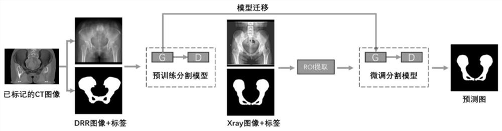
[0057] A method for establishing a hip joint segmentation model using small sample training, such as figure 2 As shown, it includes: pre-training dataset construction step, segmentation model pre-training step and segmentation model fine-tuning step.

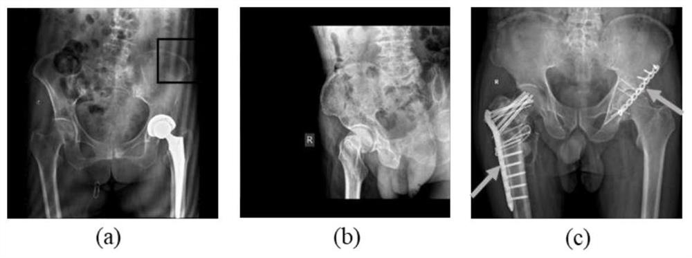
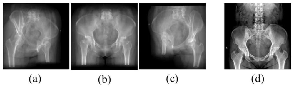
[0058] Digitally Reconstructed Radiograph (DRR) is widely used in CT simulation positioning, image-guided radiotherapy, and computer-assisted surgery. Compared with real X-ray images, the boundaries of bone tissue in CT images are more obvious. Using traditional algorithms and manual fine-tuning can get more accurate labeling data. Therefore, using a small amount of labeled CT data and using DRR projection A large number of simulated images with markers can be obtained; as an optional implementation manner, in this embodiment, a commonly used ray casting method is used to implement DRR projection. (a) to (c) in the figure are the DRR images obtained by projecting CT from multiple angles using the ray projection method. image...

Embodiment 2

[0091] A hip joint segmentation method, comprising:

[0092] Input the X-ray image to be segmented into the target detection model in the above-mentioned embodiment 1, i.e. Yolo V3, the target frame where the hip joint area is output by the target detection model;

[0093] Input the target frame where the hip joint area output by the target detection model is located into the hip joint segmentation model established by the hip joint segmentation model establishment method using small sample training provided by the above-mentioned embodiment 1, and the hip joint area is extracted by the hip joint segmentation model .

Embodiment 3

[0095] A method for establishing a hip joint segmentation model using small sample training, this embodiment is similar to the above-mentioned embodiment 1, the difference is that in the segmentation model fine-tuning step of this embodiment, no target detection is performed on real X-ray images , but directly use the small sample X-ray images of the hip joint area as the training data set.


PUM


Abstract

The invention discloses a hip joint segmentation model building method using small sample image training and application thereof, and belongs to the medical image processing field. The method comprises the following steps: projecting small sample CT data marked with a hip joint area from different angles through a digital reconstruction radiographic image method to obtain a lot of analog images with marks; forming a pre-training data set by the analog image and the hip joint area marked in the analog image; establishing a segmentation model for performing image segmentation on the input image to extract a hip joint area, and pre-training the segmentation model by using the pre-training data set; and constructing a training data set by using the small sample Xray image marked with the hip joint area, training the pre-trained segmentation model to finely adjust the segmentation model, and taking the segmentation model as the established hip joint segmentation model after the training is finished. The technical problem that an existing hip joint segmentation method is poor in segmentation result due to the fact that the existing hip joint segmentation method depends on a large amount of annotation data can be solved.

Description

technical field [0001] The invention belongs to the field of medical image processing, and more specifically relates to a method for establishing a hip joint segmentation model trained with small sample images and its application. Background technique [0002] Image segmentation is a very important technology in the field of medical image processing. The process is to extract the region of interest in the medical image. The segmentation results are useful for preoperative surgical planning, intraoperative navigation, and postoperative evaluation. important reference value. In the process of robot-assisted hip replacement surgery, the hip joint is usually used as the target area, and the 2D X-ray images acquired during the operation are registered with the 3D CT images before the operation, so as to achieve the purpose of real-time intraoperative navigation. Therefore, how to quickly and accurately segment the hip joint (pelvis, femur) from the X-ray image is a crucial part ...

Claims


Application Information

Patent Timeline
Patent Type & Authority Applications(China)
IPC IPC(8): G06T7/11G06N3/04G06N3/08G06T7/187
CPCG06N3/04G06N3/08G06T2207/30008G06T7/11G06T7/187
Inventor 李强吕进鑫梁愿怀
Owner HUAZHONG UNIV OF SCI & TECH
Why Eureka
Try Eureka