CT image three-dimensional reconstruction method based on MC-T algorithm

A CT image, MC-T technology, applied in the field of image processing, can solve the problem of unable to extract multi-threshold organs at one time, affecting the reconstruction effect, etc.

Active Publication Date: 2021-05-14
CHONGQING UNIV OF POSTS & TELECOMM
View PDF4 Cites 1 Cited by
  • Summary
  • Abstract
  • Description
  • Claims
  • Application Information

AI Technical Summary

Problems solved by technology

Aiming at the problem that the MC algorithm cannot extract multi-threshold organs at one time, Xiao Hongxu proposed a labeled 3D reconstruction algorithm. However, in the case of multiple thresholds, there is an optimal problem in the selection of label thresholds, which affects the reconstruction effect.

Method used


Image

  • CT image three-dimensional reconstruction method based on MC-T algorithm
  • CT image three-dimensional reconstruction method based on MC-T algorithm
  • CT image three-dimensional reconstruction method based on MC-T algorithm

Examples


Embodiment Construction

[0052] The following will clearly and completely describe the technical solutions in the embodiments of the present invention with reference to the accompanying drawings in the embodiments of the present invention. Obviously, the described embodiments are only some, not all, embodiments of the present invention. Based on the embodiments of the present invention, all other embodiments obtained by persons of ordinary skill in the art without making creative efforts belong to the protection scope of the present invention.

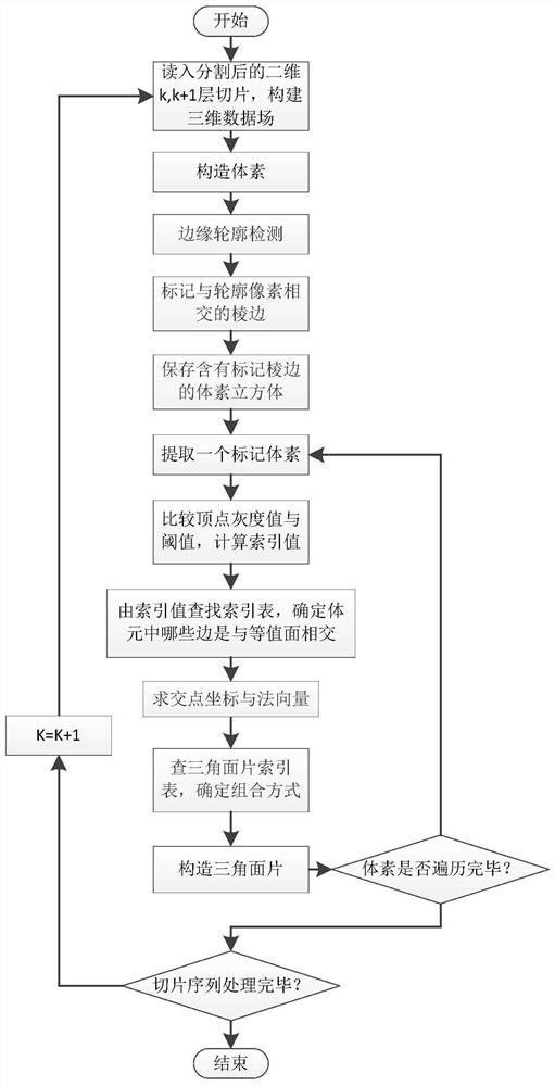
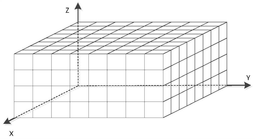
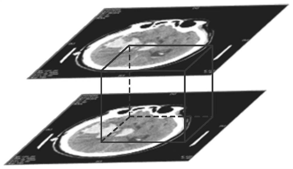
[0053] A method for three-dimensional reconstruction of CT images based on the MC-T algorithm, the method comprising: obtaining a CT image of cerebral hemorrhage to be reconstructed and a mask map of CT lesions of cerebral hemorrhage; Preprocessing: MC-T algorithm is used to reconstruct the preprocessed image data to obtain a reconstructed 3D image.

[0054] The preprocessing of the acquired data includes: segmenting the CT image of cerebral hemorrhage and CT ...


PUM


Abstract

The invention belongs to the field of image processing, and particularly relates to a CT image three-dimensional reconstruction method based on an MC-T algorithm, and the method comprises the steps: obtaining a to-be-reconstructed cerebral hemorrhage CT image and a cerebral hemorrhage CT focus mask image; preprocessing the obtained cerebral hemorrhage CT image and the cerebral hemorrhage CT focus mask image; reconstructing the preprocessed image data by adopting an MC-T algorithm to obtain a reconstructed three-dimensional image; aiming at the problems of low calculation speed and ambiguity in the traditional MC algorithm, the invention provides an improved three-dimensional reconstruction algorithm based on the MC algorithm, the algorithm takes a two-dimensional cerebral hemorrhage CT image and a focus segmentation mask slice as input, and detection of empty voxels is effectively eliminated through a voxel edge marking method in a three-dimensional data field.

Description

technical field [0001] The invention belongs to the field of image processing, and in particular relates to a method for three-dimensional reconstruction of a CT image based on an MC-T algorithm. Background technique [0002] The three-dimensional reconstruction of medical images refers to the use of two-dimensional slice images, such as sequential images such as CT and MRI, to draw models of 3D images. Common 3D reconstruction methods for medical images include volume rendering and surface rendering; volume rendering is to directly draw 3D data into a 3D model based on a certain physical model. The rendering method to reconstruct the 3D model can show more detailed information, but it requires more computer memory, and the rendering speed is slower; surface rendering uses various surface elements (triangles or quadrilaterals, etc.), and connects them through algorithms to construct them, etc. The value-surface approach is used to approximate the surface contours of real or...

Claims


Application Information

Patent Timeline
Patent Type & Authority Applications(China)
IPC IPC(8): G06T17/00G06T5/00G06T7/13G06T7/136G06T7/155
CPCG06T17/00G06T7/13G06T7/136G06T7/155G06T5/002G06T2210/41G06T2207/10081G06T2207/30016G06T2207/30096G06T2207/30204
Inventor 胡敏周秀东黄宏程
Owner CHONGQING UNIV OF POSTS & TELECOMM
Why Eureka
Try Eureka