
Gastrointestinal endoscope polyp removal collector

A collector, polyp technology, applied in the fields of suction equipment, medical science, surgery, etc., can solve the problems of difficulty in grasping, patient pain, accidental inhalation of recovery bottles, etc.

Pending Publication Date: 2021-06-15
HEBI CITY PEOPLES HOSPITAL
View PDF5 Cites 2 Cited by
  • Summary
  • Abstract
  • Description
  • Claims
  • Application Information

AI Technical Summary

Problems solved by technology

[0002] When the gastrointestinal endoscope removes polyps, it needs to be taken out by special grasping forceps or sucked on the mirror surface by negative pressure, and withdrawn with the gastrointestinal endoscope; partially removed polyps slide into the intestinal cavity, which is difficult to grasp, and the doctor gives up grasping and asks the patient to pay attention Excreted with the stool, and then sent for biopsy; some small polyps are easily inhaled into the recovery bottle by negative pressure after resection, and cannot be separated for inspection; for multiple polyps, resected under the gastroenteroscope, it is necessary to grab the polyps and exit the endoscope Afterwards, he went into the mirror again, looked for the second one, cut it again, and repeatedly entered and exited. The patient suffered a lot, and the operation was delayed, which caused a lot of inconvenience. In addition, the polyp specimen was easily contaminated by other tissue fluids, which affected the quality of the specimen, which in turn affected clinical diagnosis and effective treatment. The market Most polyp collectors collect all polyps mixed together, it is difficult to collect polyps separately according to the growth site of polyps, which affects the pathological evaluation of patients

Method used


Image

  • Gastrointestinal endoscope polyp removal collector
  • Gastrointestinal endoscope polyp removal collector

Examples


Embodiment Construction

[0015] The present invention will be described in further detail below in conjunction with the accompanying drawings.

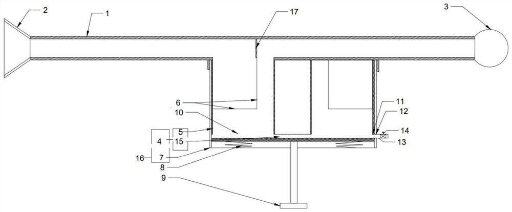
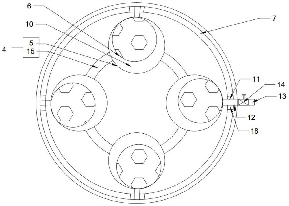
[0016] combined with Figure 1-2 As shown, a polyp collector for gastrointestinal endoscopy includes a transparent tube 1, and the two ends of the transparent tube 1 are respectively connected with an absorption cover 2 and a negative pressure device 3; the transparent tube 1 is located in the absorption cover 2 Connect with the negative pressure device 3 and be provided with collecting device 16, described collecting device 16 comprises inner tube 4, and described inner tube 4 is connected with a plurality of collecting tubes 5, and described collecting tube 5 is provided with and described transparent The filter screen 6 matched with the tube 1, the inner tube 4 is rotatably connected with an outer tube 7, the outer wall of the inner tube 4 matches the inner wall of the outer tube 7, and the bottom of the inner tube 4 is connected to the outer tube by a spr...


PUM


Abstract

The invention discloses a gastrointestinal endoscope polyp removal collector. The collector comprises a transparent tube, wherein the two ends of the transparent tube are connected with an absorption cover and a negative pressure device respectively, the transparent tube is connected with a collecting device between the absorption cover and the negative pressure device, the collecting device comprises an inner tube, a plurality of collecting tubes are connected to the interior of the inner tube, filter screens matched with the transparent tube are arranged in the collecting tubes, an outer tube is rotationally connected to the outside the inner tube, the outer wall of the inner tube is matched with the inner wall of the outer tube, the bottom of the inner tube is connected with the inner bottom wall of the outer tube through a spring, and a handle extending out of the outer tube is downwards arranged at the bottom of the inner tube. Compared with the prior art, the collector has the advantages that the structure is simple, polyps can be rapidly removed, and the polyps can be filtered and collected respectively.

Description

technical field [0001] The invention relates to the technical field of medical devices, in particular to a polyp collector for gastrointestinal endoscopy. Background technique [0002] When the gastrointestinal endoscope removes polyps, it needs to be taken out by special grasping forceps or sucked on the mirror surface by negative pressure, and withdrawn with the gastrointestinal endoscope; partially removed polyps slide into the intestinal cavity, which is difficult to grasp, and the doctor gives up grasping and asks the patient to pay attention Excreted with the stool, and then sent for biopsy; some small polyps are easily inhaled into the recovery bottle by negative pressure after resection, and cannot be separated for inspection; for multiple polyps, resected under the gastroenteroscope, it is necessary to grab the polyps and exit the endoscope Afterwards, he went into the mirror again, looked for the second one, cut it again, and repeatedly entered and exited. The pati...

Claims


Application Information

Patent Timeline
IPC IPC(8): A61B17/00A61M1/00
CPCA61B17/00234A61B2217/002A61B2217/005
Inventor 申利敏吴怡晨闫晓云
Owner HEBI CITY PEOPLES HOSPITAL
Learn More