Multi-modal brain image data processing method and device, equipment and storage medium
A data processing and brain imaging technology, applied in character and pattern recognition, recognition of medical/anatomical patterns, instruments, etc., can solve the problems of ineffective use of complementary information and high reliability of biomarkers
- Summary
- Abstract
- Description
- Claims
- Application Information
AI Technical Summary
Problems solved by technology
Method used
Image
Examples
Embodiment 1
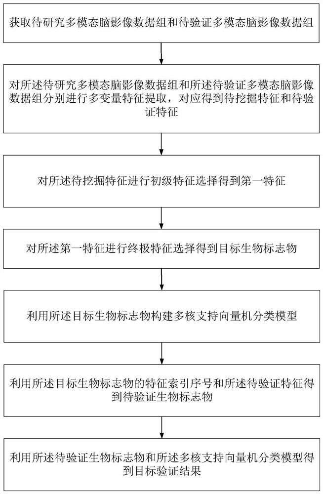
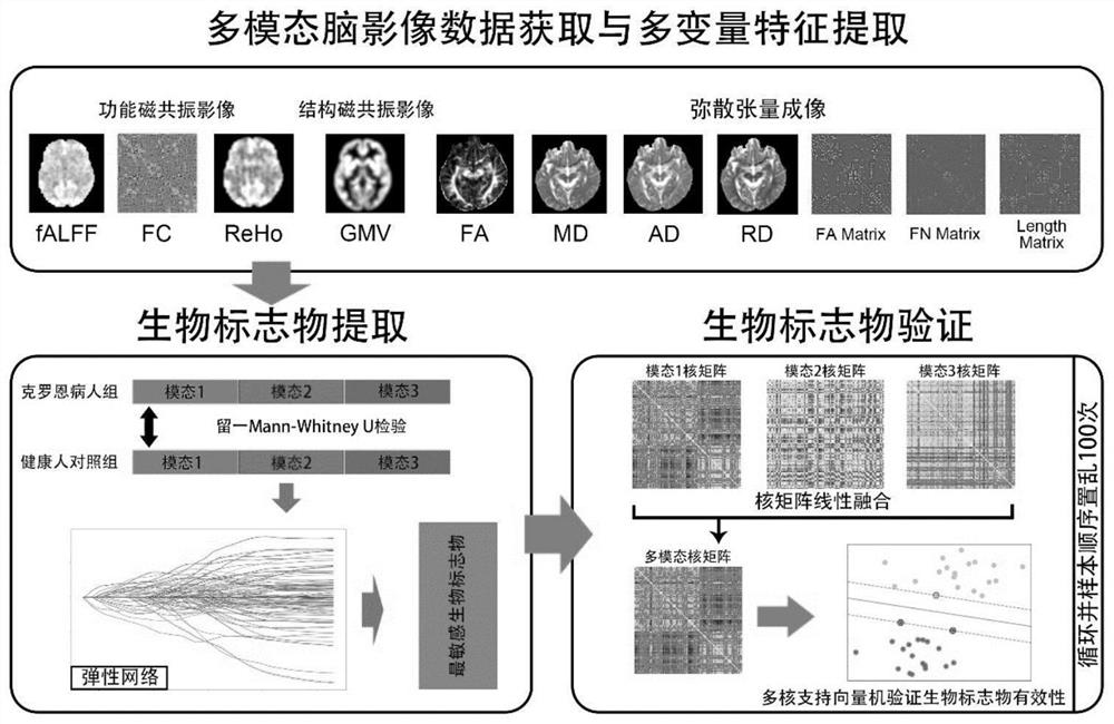
[0051] See figure 1 and figure 2 , figure 1 It is a flow chart of a method for processing multimodal brain image data provided in this embodiment, figure 2 It is a schematic diagram of a method for processing multimodal brain image data provided in this embodiment. This embodiment discloses a method for processing multimodal brain image data, including:
[0052] Step 1. Obtain the multimodal brain imaging data set to be studied and the multimodal brain imaging data set to be verified.
[0053] In this embodiment, firstly, it is necessary to obtain the multimodal brain image data set to be studied and the multimodal brain image data set to be verified, and both the multimodal brain image data set to be studied and the multimodal brain image data set to be verified Including: resting state functional magnetic resonance imaging modality data set, diffusion tensor imaging modality data set and structural magnetic resonance imaging modality data set.
[0054] Specifically, the...
Embodiment 2
[0112] See Figure 5 , Figure 5 It is a schematic structural diagram of a device for multimodal brain image data processing provided in this embodiment. This embodiment discloses a device for processing multimodal brain image data, including:
[0113] Image acquisition module 1, used to acquire multimodal brain imaging data to be studied and multimodal brain imaging data to be verified;
[0114] The feature extraction module 2 is used to perform multivariate feature extraction on the multi-modal brain image data to be studied and the multi-modal brain image data to be verified, and correspondingly obtain the features to be mined and the features to be verified;
[0115] The biomarker extraction module 3 is used to perform feature selection on the features to be mined to obtain the target biomarkers, use the target biomarkers to construct a multi-core support vector machine classification model, and use the target biomarkers and the features to be verified to obtain the biom...
Embodiment 3
[0122] See Figure 6 , Figure 6 It is a schematic structural diagram of a device for processing multimodal brain image data provided in this embodiment. This embodiment discloses a device for processing multimodal brain image data, including: a processor, a communication interface, a memory, and a communication bus, wherein the processor, the communication interface, and the memory complete communication with each other through the communication bus;
[0123] memory for storing computer programs;
[0124] The processor is configured to implement the method steps in any one of the present embodiments when executing the computer program.
[0125] The device for processing multi-modal brain image data provided by the embodiment of the present invention can execute the above-mentioned method embodiment, and its implementation principle and technical effect are similar, and will not be repeated here.
PUM
 Abstract
Description
Claims
Application Information
 - R&D
- Intellectual Property
- Life Sciences
- Materials
- Tech Scout
- Unparalleled Data Quality
- Higher Quality Content
- 60% Fewer Hallucinations
Browse by: Latest US Patents, China's latest patents, Technical Efficacy Thesaurus, Application Domain, Technology Topic, Popular Technical Reports.
© 2025 PatSnap. All rights reserved.Legal|Privacy policy|Modern Slavery Act Transparency Statement|Sitemap|About US| Contact US: help@patsnap.com


