
Section tissue identification method and device, cell identification model and tissue segmentation model

A cell identification and tissue identification technology, applied in the field of image processing, can solve problems such as low efficiency and increase the workload of pathologists, and achieve the effect of maintaining integrity

Pending Publication Date: 2021-07-02
广州信瑞医疗技术有限公司
View PDF0 Cites 6 Cited by
  • Summary
  • Abstract
  • Description
  • Claims
  • Application Information

AI Technical Summary

Problems solved by technology

[0004] When a doctor performs pathological screening on a patient, it is usually necessary to check the pathological section, to confirm the observed tissue and the cells in the tissue, and to make an accurate diagnosis of the patient's symptoms through empirical judgment. Diagnosis, this method seriously increases the workload of pathologists, and the efficiency of this method is too low

Method used


Image

  • Section tissue identification method and device, cell identification model and tissue segmentation model
  • Section tissue identification method and device, cell identification model and tissue segmentation model
  • Section tissue identification method and device, cell identification model and tissue segmentation model

Examples


Embodiment Construction

[0033] The technical solutions in the embodiments of the present application will be described below with reference to the drawings in the embodiments of the present application.

[0034] In view of the fact that when doctors do pathological screening of patients, they usually need to check the pathological slides, confirm the observed tissue and the cells in the tissue, and make an accurate diagnosis of the patient's symptoms through empirical judgment. Diagnosis, this method seriously increases the workload of pathologists, and the efficiency of this method is too low. After research and exploration, the inventors of the present application propose the following embodiments to solve the above problems.

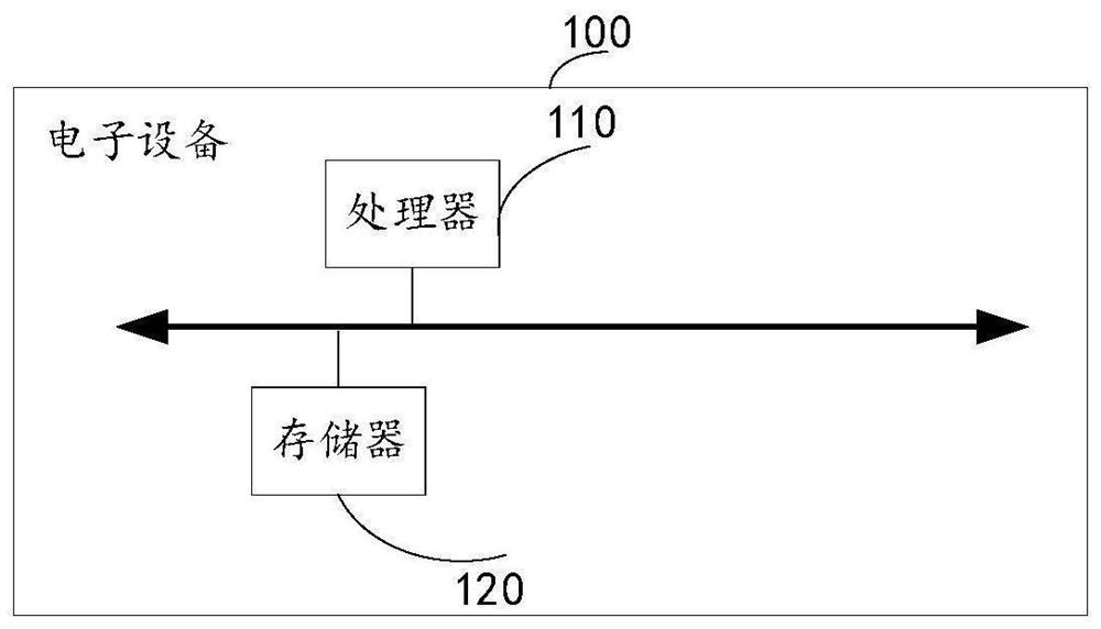
[0035] see figure 1 , a schematic structural block diagram of an electronic device 100 applying the sliced ​​tissue identification method and apparatus provided in the embodiment of the present application. In the embodiment of the present application, the electronic device...


PUM


Abstract

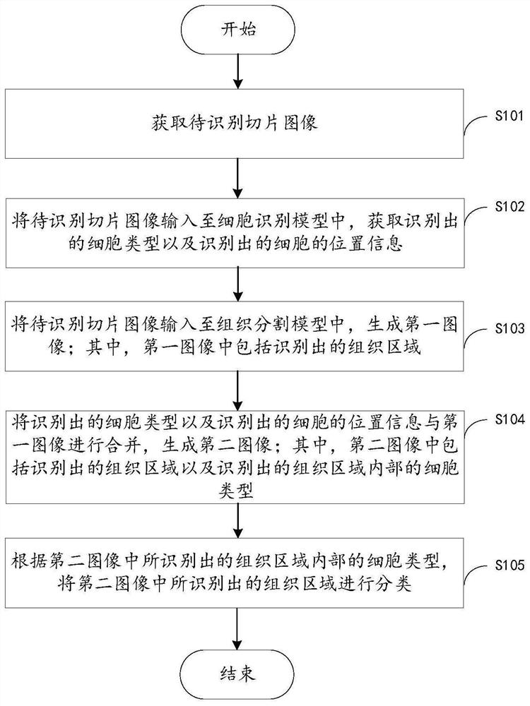
The invention provides a slice tissue identification method and device, a cell identification model and a tissue segmentation model. The method comprises the following steps: acquiring a to-be-identified slice image; inputting the to-be-identified slice image into a cell identification model, and obtaining the identified cell type and the identified cell position information; inputting the to-be-identified slice image into the tissue segmentation model to generate a first image; wherein the first image comprises an identified tissue area; combining the identified cell type and the identified cell position information with the first image to generate a second image; wherein the second image comprises the identified tissue area and the cell type in the identified tissue area; and classifying the tissue areas identified in the second image according to the cell types in the tissue areas identified in the second image. Through the above mode, the workload of a pathologist is reduced, and the identification efficiency and the identification accuracy of the slice tissue are improved.

Description

technical field [0001] The present application relates to the technical field of image processing, in particular, to a sliced ​​tissue recognition method and device, a cell recognition model and a tissue segmentation model. Background technique [0002] A pathological section refers to a thin section made of pathological tissue of a certain size and made by histopathological methods. When making it, the diseased tissue is usually embedded in a paraffin block, sliced ​​into thin slices with a microtome, stained, and further examined with a microscope. Doctors need to use a high-power microscope to review images, check the pathological changes of tissue or cells in the specimen, explore the cause, pathogenesis and process of the lesion, and finally make a pathological diagnosis. Histopathology is different from cytopathology. Histopathology often depends on the distribution, quantity, and cell characteristics of one or more types of cells in different tissue regions when diag...

Claims


Application Information

Patent Timeline
IPC IPC(8): G06T7/00G06T7/11G06K9/32G06K9/38G06K9/62G06N3/04G06N3/08
CPCG06T7/0012G06T7/11G06N3/08G06T2207/10056G06T2207/10024G06T2207/20081G06T2207/20084G06T2207/30024G06V10/25G06V10/28G06N3/048G06N3/045G06F18/241
Inventor 周丕轩李志宁郝昀超张国根
Owner 广州信瑞医疗技术有限公司
Learn More