A component, system and application for quantitative detection of prostate cancer biomarker miRNA-141
A technology of miRNA-141 and biomarkers, which is applied in the determination/inspection of microorganisms, biochemical equipment and methods, etc., can solve the problems of miRNA blood background complex, complex and cumbersome process, etc., to achieve improved sensitivity, reliable analysis results, and convenient The effect of carrying and transporting
- Summary
- Abstract
- Description
- Claims
- Application Information
AI Technical Summary
Problems solved by technology
Method used
Image
Examples
Embodiment 1
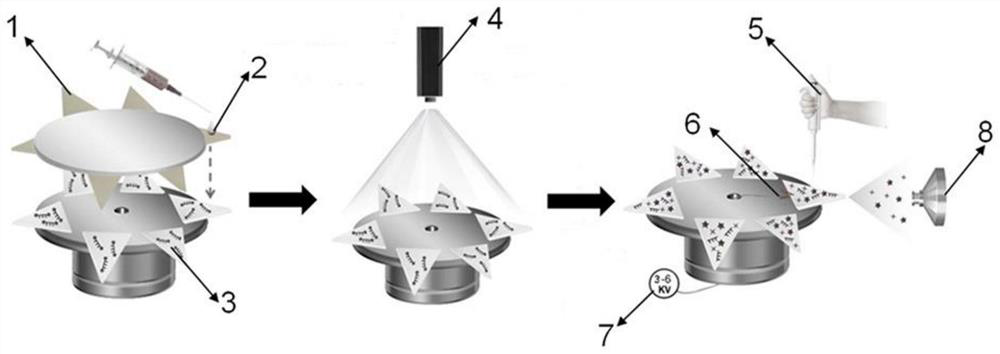
[0059] The preparation method of paper base, comprises the steps:
[0060] Gold nanoparticles were synthesized by the Turk evich method. Trisodium citrate reduction of HAuCl 4 ·3H 2 O to synthesize gold nanoparticles of citrate. The specific synthesis steps are as follows, 94mL of HAuCl 4 The aqueous solution (2 mM) was heated to 110°C, stirred until boiling vigorously, and then 19 mL of trisodium citrate solution (38.8 mM) was added immediately. The mixture was kept boiling for a certain time until the color of the solution gradually changed from yellow to wine red. The reaction was terminated when the suspension was cooled to room temperature. The concentration of as-synthesized gold nanoparticles was calculated to be 6 × 10 -8 M, and stored at 4°C for further use.
[0061] A PCs-sRNA probe containing gold nanoparticles and DNA strands was synthesized, wherein the gold nanoparticles were able to form Au-S bonds with the thiol-modified DNA strand SH-L, thus realizing t...
Embodiment 2
[0068] The specific steps to verify the strand displacement reaction are as follows (experimental results such as figure 2 shown):
[0069] (1) Prepare thiolated DNA strand (SH-L), signal strand (PCs-sRNA), SH-L (3.0 μM) and PCs-sRNA (6.2 μM) in 20 mM Tris-HCl buffer (100 mM Na + , pH=7.4) was heated to 90°C for 5 min and then cooled to room temperature for 2 hours to form a ternary DNA duplex probe. Based on Marker, SH-L, PCs-sRNA and ternary DNA were verified by PAGE gel. This step verifies the successful binding of ternary DNA double strands.
[0070] (2) Divide the remaining ternary DNA solution into two parts, add miRNA-141 (50 pM) to one solution, add an equal volume of water to the other solution, heat the two mixed solutions to 90°C for 5 min, and then cool them to 2 hours at room temperature. Based on Marker, SH-L, PCs-sRNA, miRNA-141, the mixed solution with water added in this step, and the mixed solution with miRNA-141 were used for gel running verification. ...
Embodiment 3
[0074] At 37°C, PCs-AuNPs nanoprobes were added dropwise to the thiol-modified paper substrate, then different concentrations of miRNA-141 (18 μL) were added dropwise and incubated for 60 min, and F chain (1 μM, 18 μL) was added dropwise and incubated for the same time. During , different matrix solutions (here, serum matrix solution and pH=7.4 ammonium acetate buffer solution) were used to keep the paper substrate wet, and then infiltrated into the lower paper substrate for paper spray mass spectrometry detection. Take lg c as the abscissa and the mass spectrum peak intensity ratio of the signal molecule to the internal standard as the ordinate to draw a standard curve, the specific concentrations are 50fM, 500fM, 1pM, 5pM, 10pM, 50pM, the results are as follows Figure 4 As shown, it can be seen that the method of the present invention has a good linear relationship in quantitative analysis.
PUM
 Abstract
Description
Claims
Application Information
 - R&D
- Intellectual Property
- Life Sciences
- Materials
- Tech Scout
- Unparalleled Data Quality
- Higher Quality Content
- 60% Fewer Hallucinations
Browse by: Latest US Patents, China's latest patents, Technical Efficacy Thesaurus, Application Domain, Technology Topic, Popular Technical Reports.
© 2025 PatSnap. All rights reserved.Legal|Privacy policy|Modern Slavery Act Transparency Statement|Sitemap|About US| Contact US: help@patsnap.com


