Construction method of brain corpus callosum segmentation prediction image for corpus callosum state evaluation

A technology for predicting images and constructing methods, which is applied in the field of medical image segmentation and deep learning, and can solve problems such as high error rate, low detection rate of fetal corpus callosum abnormalities, and inability to accurately calculate the volume of corpus callosum

Active Publication Date: 2021-07-23
WEST CHINA HOSPITAL SICHUAN UNIV
View PDF14 Cites 5 Cited by
  • Summary
  • Abstract
  • Description
  • Claims
  • Application Information

AI Technical Summary

Problems solved by technology

[0003] At present, the measurement of the size and position of the corpus callosum still relies on medical imaging technicians to provide it according to the fetal brain ultrasound image annotation, which requires high requirements for the operator's experience and tech

Method used


Image

  • Construction method of brain corpus callosum segmentation prediction image for corpus callosum state evaluation
  • Construction method of brain corpus callosum segmentation prediction image for corpus callosum state evaluation
  • Construction method of brain corpus callosum segmentation prediction image for corpus callosum state evaluation

Examples


Embodiment 1

[0036] Embodiment 1 The construction of the fetal ultrasound image state analysis deep neural network model of the present invention

[0037] (1) Image preprocessing

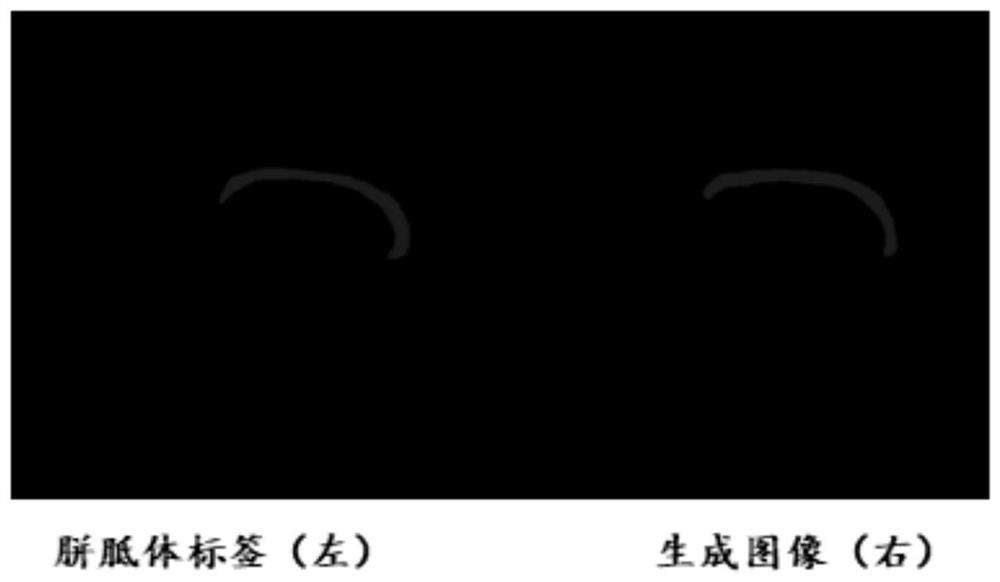
[0038] a. Acquisition of ultrasound images of the fetal brain and segmentation label images of the corpus callosum

[0039] Ultrasound images of the fetal brain were collected by the TRT33 frequency-variable dual-plane brain probe using brightness modulation type ultrasonic section imager; the segmentation label image of the corpus callosum of the brain was provided by medical imaging technicians according to the fetal brain ultrasound image annotation;

[0040] b. Image data preprocessing

[0041]The acquired ultrasound images of the fetal brain are translated, transformed, twisted and enhanced, and elastically deformed, and the corner point detection and center point detection are performed on the corpus callosum segmentation label image of the brain. The specific method is as follows:

[0042] ① Use the hor...

Embodiment 2

[0069] Embodiment 2 Fetal ultrasound image state analysis of the present invention

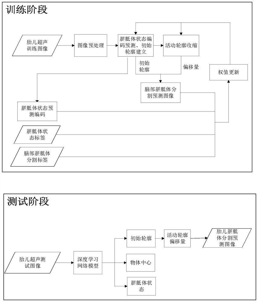
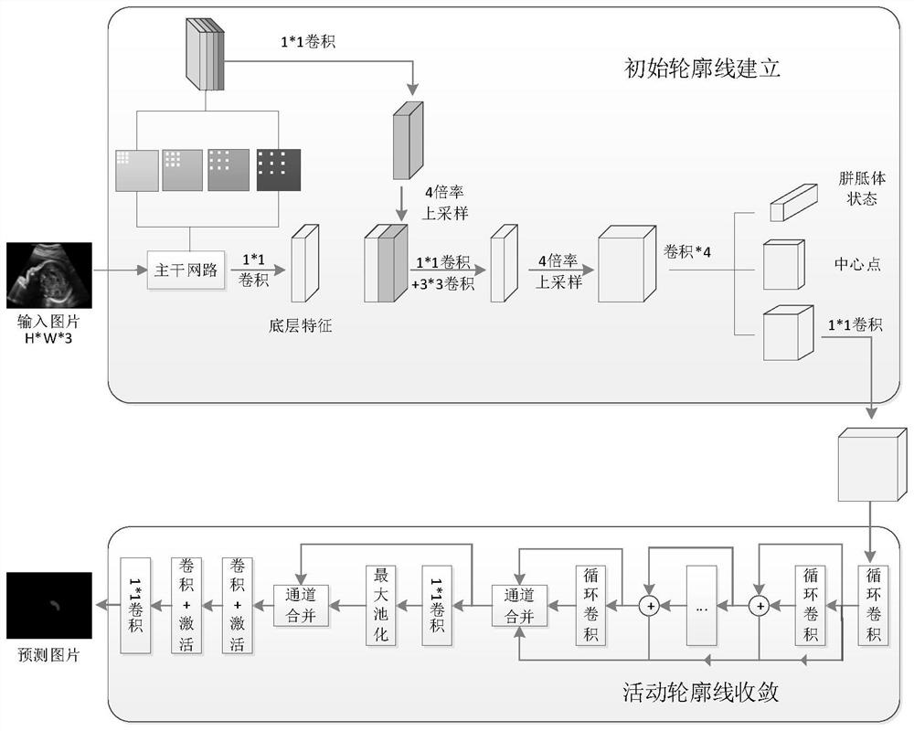
[0070] Take a fetal brain ultrasound image data to be evaluated and input it into the deep neural network model constructed in Example 1, and construct a brain corpus callosum segmentation map through the output initial contour line and active contour offset to evaluate the state of the fetal brain corpus callosum , for the specific frame structure of corpus callosum state analysis based on deep neural network fetal ultrasound images, see image 3 .

[0071] In summary, the present invention converts the segmentation of the corpus callosum into the initial contour establishment and active contour convergence, uses the encoding and decoding module to obtain multi-scale image feature information, predicts the corpus callosum state code and initial contour of the fetal ultrasound image, and distributes the vector through the key points The construction of the sum loss function makes the key poin...


PUM


Abstract

The invention discloses a construction method of a brain corpus callosum segmentation prediction image for corpus callosum state evaluation, which comprises the following steps: drawing a corpus callosum initial contour line from an acquired fetal brain ultrasonic image, and calculating a key point offset of the corpus callosum initial contour line; and cutting a brain corpus callosum segmentation prediction image from the fetal brain ultrasound image according to the initial contour line of the corpus callosum and the key point offset. According to the deep neural network model for fetal ultrasound image state analysis, the blank of brain ultrasound image brain corpus callosum state analysis is filled, and a method for evaluating the brain corpus callosum state through a brain ultrasound image is created for the first time.

Description

technical field [0001] The invention relates to the field of medical image segmentation and deep learning, and specifically relates to a method for constructing a predictive image of brain corpus callosum segmentation for corpus callosum state assessment. Background technique [0002] The corpus callosum is located at the floor of the interhemispheric fissure and is the largest commissural fiber in the cerebral hemisphere. Dysplasia of the corpus callosum (Agenesis of Corpus Callosum, ACC) is a congenital abnormality in fetal central nervous system malformation, which refers to the partial or complete loss of the corpus callosum during development. It is important to use imaging examinations to diagnose the development of the corpus callosum during fetal life. effect. [0003] At present, the measurement of the size and position of the corpus callosum still relies on medical imaging technicians to provide it according to the fetal brain ultrasound image annotation, which re...

Claims


Application Information

Patent Timeline
IPC IPC(8): G06T7/187G06T7/11G06N3/04G06N3/08G16H30/20
CPCG06T7/187G06T7/11G06N3/08G16H30/20G06T2207/20221G06T2207/20016G06N3/045
Inventor 曹桂群何长涛程建陈玉兰郑文刘鑫周柱玉宋思思
Owner WEST CHINA HOSPITAL SICHUAN UNIV
Why Eureka
Try Eureka