Mathematical algorithm microscopic bladder endoscope imaging system and image processing method

An image processing and mathematical algorithm technology, applied in the field of medical imaging, can solve problems such as poor image quality, lack of tumor risk assessment, unfavorable diagnosis, etc., to achieve a clearer image, facilitate accurate observation and diagnosis, and improve the effect of denoising performance

Pending Publication Date: 2021-09-21
韩从辉
View PDF0 Cites 0 Cited by
  • Summary
  • Abstract
  • Description
  • Claims
  • Application Information

AI Technical Summary

Problems solved by technology

However, the endoscopic system in the prior art lacks the processing of judging the risk of tumors based on medical images, and the quality of the collected images is often poor, which is not conducive to diagnosis

Method used


Image

  • Mathematical algorithm microscopic bladder endoscope imaging system and image processing method
  • Mathematical algorithm microscopic bladder endoscope imaging system and image processing method
  • Mathematical algorithm microscopic bladder endoscope imaging system and image processing method

Examples


Embodiment

[0028] Embodiment Mathematical Algorithm Microbladder Endoscope Imaging System and Image Processing Method

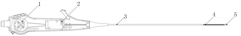
[0029] A mathematical algorithm microbladder endoscope imaging system, including an endoscope handle, an outer sheath 3, an image processing module and a display module, the structure diagram of the endoscope handle and the outer sheath 3 is as follows figure 1 As shown, the endoscope handle is provided with a direction control mechanism 1, a light source module and an image receiving module 2, one end of the endoscope handle is connected to one end of the outer sheath tube 3, and the other end of the outer sheath tube 3 is provided with a bending part 4 The connecting end of the outer sheath tube 3 and the bending part 4 is a composite braided tube, and the bending part 4 is a two-way serpentine tube, which can realize a two-way deflection of 270°. The direction control mechanism 1 is connected with the bending part 4 to control the bending part 4. In the bending direc...


PUM


Abstract

The invention belongs to the technical field of medical imaging, and discloses a mathematical algorithm microscopic bladder endoscope imaging system and an image processing method. The system comprises an endoscope handle, an outer sheath tube, an image processing module and a display module; the endoscope handle is connected with the outer sheath tube, the outer sheath tube is provided with a bending part, a direction control mechanism is connected with the bending part, and the direction control mechanism is connected with the image processing module; the end, away from the outer sheath tube, of the bent portion is provided with a tip portion, an image sensor is arranged in the tip portion, the light source module provides a light source for the image sensor, the image sensor is connected with the image receiving module, the image receiving module is connected with the image processing module, and the image processing module is connected with the display module. The method comprises the steps that the light source module provides a light source for the image sensor, controls the angle of the bending part, collects an image in the body of a patient, carries out image processing, and displays image information in the body of the patient on the display module. According to the invention, the quality of medical images is improved, and research and judgment of tumor risks are realized. The device is suitable for medical staff to observe conditions in the body of a patient.

Description

technical field [0001] The invention belongs to the technical field of medical imaging and relates to an endoscope, in particular to a mathematical algorithm microbladder endoscope imaging system and an image processing method. Background technique [0002] At present, for the early diagnosis of tumors, the commonly used clinical methods mainly include: X-ray examination, electron microscope examination, radionuclide examination, cytology examination, surgical examination, ECT examination, etc., but all of them can only achieve middle and late diagnosis. For most cancers, only when the number of cancer cells reaches 1 billion can the tumor diameter reach about 1 cm. However, current imaging diagnostic techniques such as CT and PET have a minimum positive resolution of only 0.6-1 cm. When the tumor grows to about 2 cm, Histological pathological examination can be carried out, which brings great difficulties to the early clinical diagnosis of cancer. [0003] The traditional ...

Claims


Application Information

Patent Timeline
IPC IPC(8): G06T7/00G06T5/00G06K9/62G06T7/90G16H30/20G16H50/30
CPCG06T7/0012G16H30/20G16H50/30G06T7/90G06T2207/10068G06T2207/30096G06F18/29G06T5/73G06T5/70
Inventor 韩从辉史振铎
Owner 韩从辉
Why Eureka
Try Eureka