A ray-casting-based region-of-interest fusion method for bimodal volume data

A technology of region of interest and fusion method, which is applied in the field of 3D volume data visualization, can solve problems that are rarely considered, achieve good fusion, and improve the efficiency of visual analysis

Active Publication Date: 2022-07-12
BEIJING INSTITUTE OF TECHNOLOGYGY
View PDF2 Cites 0 Cited by
  • Summary
  • Abstract
  • Description
  • Claims
  • Application Information

AI Technical Summary

Problems solved by technology

Because the fusion process only considers the fusion of the attributes of the corresponding sample points, there is no or little consideration of the influence of other sample points and voxels around the sampling point on the projection ray

Method used


Image

  • A ray-casting-based region-of-interest fusion method for bimodal volume data
  • A ray-casting-based region-of-interest fusion method for bimodal volume data
  • A ray-casting-based region-of-interest fusion method for bimodal volume data

Examples


Embodiment Construction

[0046] The specific embodiments of the present invention will be described below with reference to the accompanying drawings and examples.

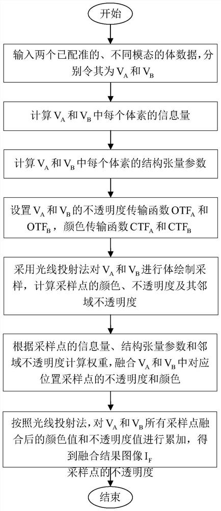
[0047] figure 1 A flow chart of a ray-casting-based bimodal volume data region-of-interest fusion method according to the present invention is given, and its main steps are as follows:

[0048] Step S1: Input two registered volume data of different modalities, and let them be V A and V B .

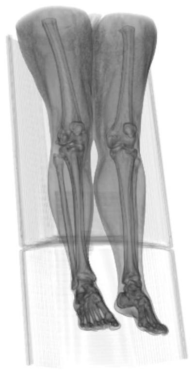
[0049] The modalities fused in this example include CT and PET, and the data used are from the cancer public image database TCIA (Clark K, Vendt B, Smith K, et al. The Cancer ImagingArchive (TCIA): maintaining and operating a public information repository .Journal of DigitalIma-ging,2013,26(6):1045-1057.), is the leg scan data of a patient with moderate synovial sarcoma, the size of the data is 256×256×127. Let CT data be V A , PET data is V B .

[0050] Step S2: Calculate V separately A and V B The amount of information in each voxel is calc...


PUM


Abstract

The invention relates to a ray-casting-based dual-modal volume data region-of-interest fusion method, which belongs to the field of three-dimensional volume data visualization, and aims to reduce the influence of mutual occlusion of different modal structural features in traditional volume rendering fusion. In the case of given different modal volume data, the method first performs volume rendering sampling on the volume data according to the ray casting method, and selects the region of interest through the volume rendering transfer function; The fusion weight is calculated with the neighborhood opacity, and the sampling points of different modes are fused; finally, the fusion results of all sampling points are accumulated according to the ray casting method to obtain the fusion image. This method can alleviate the influence of the mutual occlusion of different modal structural features in multimodal volume data fusion, which is beneficial to the identification and localization of modal features.

Description

technical field [0001] The invention relates to a bimodal volume data fusion method, in particular to a ray projection-based bimodal volume data interest region fusion method, which belongs to the field of three-dimensional volume data visualization. Background technique [0002] With the update and iteration of medical equipment, many CT (Computed Tomography) and PET (PositronEmission Computed Tomography) equipment can directly collect 3D medical image information and output 3D medical volume data. Visualizing these volume data directly allows doctors to obtain the spatial structure and morphological information of tissues or organs more intuitively and completely. However, due to different imaging principles, images collected by different imaging devices have different characteristics. For example, CT images have high resolution and can clearly display dense tissue structures such as bones; PET images have low resolution, but can provide high-precision physiological and m...

Claims


Application Information

Patent Timeline
Patent Type & Authority Patents(China)
IPC IPC(8): G06T15/00G06T19/20
CPCG06T15/005G06T19/20G06T2200/04G06T2210/41
Inventor 张文耀付靖妃王成
Owner BEIJING INSTITUTE OF TECHNOLOGYGY
Why Eureka
Try Eureka