Check patentability & draft patents in minutes with Patsnap Eureka AI!

A method and system for image recognition of porcine intestinal epithelial damage repair

A technology for damage repair and image recognition, applied in image analysis, image enhancement, image data processing, etc., to reduce errors, improve recognition rate and clarity, and improve accuracy

Active Publication Date: 2021-11-16
ANIMAL SCI RES INST GUANGDONG ACADEMY OF AGRI SCI
View PDF5 Cites 0 Cited by
  • Summary
  • Abstract
  • Description
  • Claims
  • Application Information

AI Technical Summary

Problems solved by technology

[0003] The purpose of the present invention is to propose a pig intestinal epithelial injury repair image recognition method and system to solve one or more technical problems in the prior art, at least provide a beneficial choice or create conditions

Method used


Image

  • A method and system for image recognition of porcine intestinal epithelial damage repair
  • A method and system for image recognition of porcine intestinal epithelial damage repair

Examples


Embodiment Construction

[0068] The concept, specific structure and technical effects of the present disclosure will be clearly and completely described below in conjunction with the embodiments and drawings, so as to fully understand the purpose, scheme and effect of the present disclosure. It should be noted that, in the case of no conflict, the embodiments in the present application and the features in the embodiments can be combined with each other.

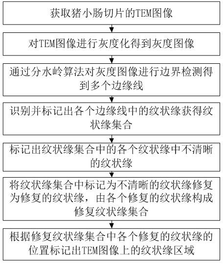
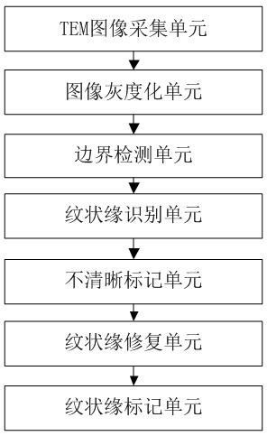
[0069] like figure 1 Shown is a flow chart of a pig intestinal epithelial injury repair image recognition method, combined below figure 1 To illustrate a method for image recognition of pig intestinal epithelial damage repair according to an embodiment of the present invention, the method includes the following steps:

[0070] S100, acquiring a TEM image of a slice of pig small intestine;

[0071] S200, grayscale the TEM image to obtain a grayscale image;

[0072] S300, performing boundary detection on the grayscale image through a watershed algorit...


PUM


Abstract

The invention discloses an image recognition method and system for pig intestinal epithelial damage repair. By obtaining a TEM image of a section of pig small intestine; The unclear striae in each striae in the striae set; the striae marked as unclear in the striae set is repaired to obtain the repaired striae set; according to each repair in the striae set The position of the striated border marks the striated border area on the TEM image, which can solve the problems of unclear boundaries between porcine intestinal epithelial cells, overlapping striated borders, and adhesion, and improve the recognition rate and clarity of striated borders , so that the follow-up statistical analysis of the striated border and the intestinal epithelial injury repair model for animal experiments constructed based on the striated border can reduce errors and improve the accuracy of statistical results.

Description

technical field [0001] The disclosure belongs to the fields of computer vision, image processing technology, and data analysis technology, and in particular relates to an image recognition method and system for repairing pig intestinal epithelium damage. Background technique [0002] At present, the intestinal system has the largest mucosal surface in the body and is composed of a single layer of intestinal epithelial cells capable of continuous self-renewal. and damage from the external environment. The general process of sampling images is to slice the small intestine of the pig, fix it with Bouin's solution, embed it in paraffin, and stain it with hematoxylin-eosin (HE staining). The finger-like protrusions formed by the longitudinal section of the villi are covered with a layer of columnar epithelial cells. On the free surface of the cells (the side facing the intestinal cavity), a thin layer of red-stained columnar cells often adheres to the surface of the cells under ...

Claims


Application Information

Patent Timeline
Patent Type & Authority Patents(China)
IPC IPC(8): G06T7/00G06T7/13
CPCG06T7/0012G06T7/13G06T2207/10056G06T2207/30028
Inventor 鲁慧杰马现永余苗田志梅容庭邓盾崔艺燕刘志昌李贞明宋敏
Owner ANIMAL SCI RES INST GUANGDONG ACADEMY OF AGRI SCI
Features
  • R&D
  • Intellectual Property
  • Life Sciences
  • Materials
  • Tech Scout
  • Unparalleled Data Quality
  • Higher Quality Content
  • 60% Fewer Hallucinations
Learn More