
X-ray chest radiograph pseudo focus image generation method and device and storage medium

An image generation device and image generation technology, applied in image enhancement, image analysis, medical image and other directions, can solve problems such as offset, inability to bring more information from network training, and poor authenticity.

Pending Publication Date: 2021-10-22
JIANGMEN POLYTECHNIC
View PDF1 Cites 1 Cited by
  • Summary
  • Abstract
  • Description
  • Claims
  • Application Information

AI Technical Summary

Problems solved by technology

However, there are deficiencies in the above two methods. Although the image generated by the first method is very real, it is very different from the source image and cannot bring more information to the network training; although the second method generates a new lesion images, whose realism is too poor compared to real lesion images, which may lead to a shift in network training

Method used


Image

  • X-ray chest radiograph pseudo focus image generation method and device and storage medium
  • X-ray chest radiograph pseudo focus image generation method and device and storage medium
  • X-ray chest radiograph pseudo focus image generation method and device and storage medium

Examples


Embodiment Construction

[0052] Embodiments of the present invention are described in detail below, examples of which are shown in the drawings, wherein the same or similar reference numerals designate the same or similar elements or elements having the same or similar functions throughout. The embodiments described below by referring to the figures are exemplary only for explaining the present invention and should not be construed as limiting the present invention.

[0053] In the description of the present invention, it should be understood that the orientation descriptions, such as up, down, front, back, left, right, etc. indicated orientations or positional relationships are based on the orientations or positional relationships shown in the drawings, and are only In order to facilitate the description of the present invention and simplify the description, it does not indicate or imply that the device or element referred to must have a specific orientation, be constructed and operated in a specific ...


PUM


Abstract

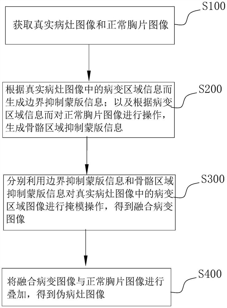
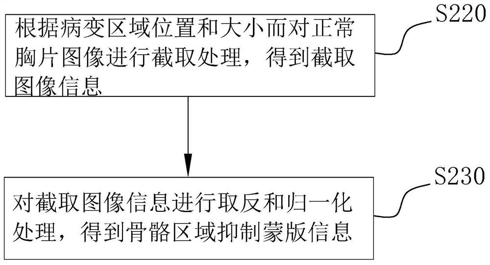
The invention discloses an X-ray chest radiograph pseudo focus image generation method and device and a storage medium. The method comprises the steps: acquiring a real focus image and a normal chest radiograph image; generating boundary suppression mask information according to lesion area information in the real lesion image; according to the lesion area information, operating the normal chest radiograph image to generate skeleton area inhibition mask information; performing mask operation on a lesion area image in the real lesion image by using the boundary inhibition mask information and the skeleton area inhibition mask information to obtain a fused lesion image; and superposing the fused lesion image and the normal chest radiograph image to obtain a pseudo lesion image. Through the technical scheme of the embodiment, more training data can be provided for deep learning, and the robustness and accuracy of a deep learning network are improved.

Description

technical field [0001] The present invention relates to the technical field of image generation, in particular to a method, device and storage medium for generating pseudo-focus images of X-ray chest films. Background technique [0002] When applying artificial intelligence methods for lung disease diagnosis, it is best to give the location of the lesion area to make the artificial intelligence diagnosis more convincing. In recent years, deep learning methods have been widely used in automatic diagnosis of medical images. However, the annotation of medical images requires expertise and a lot of time, which limits the application of deep learning techniques in medical image analysis. The existing method is to use the marked lesion images to generate more lesion images through image flipping, displacement, clipping, etc.; or to generate pseudo-abnormal images for network training by intercepting and simply pasting the lesion images. [0003] There are two types of existing p...

Claims


Application Information

Patent Timeline
Patent Type & Authority Applications(China)
IPC IPC(8): G16H30/20G06T7/70G06T7/11G06T5/50G06N3/08G06N3/04
CPCG16H30/20G06T5/50G06T7/11G06N3/08G06T7/70G06T2207/10116G06T2207/30061G06N3/045
Inventor 艾星芳郑士基杨镇首
Owner JIANGMEN POLYTECHNIC
Learn More