Percutaneous kidney ultramicro-channel visible puncture and expansion integrated kit with negative pressure suction

A technology for puncture expansion and negative pressure channels, which is applied in the direction of puncture needles, parts of surgical instruments, surgical instruments for suctioning substances, etc. Minimize the risk of surgery, ensure suction efficiency, and meet the effect of puncture

Active Publication Date: 2021-11-05
NINGBO FIRST HOSPITAL
View PDF7 Cites 3 Cited by
  • Summary
  • Abstract
  • Description
  • Claims
  • Application Information

AI Technical Summary

Problems solved by technology

Although ultra-micro-channel percutaneous nephrolithotomy (super-mini-PCNL, SMP) with a perfusion negative pressure suction system can better solve the problems of insufficient perfusion, unclear vision, and high internal pressure of the renal pelvis, SMP is only suitable for Management of kidney stones <2.5 cm in diameter
[0004] In addition, when traditional percutaneous nephroscopic surgery enters the kidney due to puncture, the operator relies on hand feeling, or predicts the depth under the guidance of ultrasound, CT, etc. to judge the puncture site. These methods are blind, time-consuming, and occasionally Mistakes can easily lead to bleeding, residual stones, sepsis and other complications

Method used


Image

  • Percutaneous kidney ultramicro-channel visible puncture and expansion integrated kit with negative pressure suction
  • Percutaneous kidney ultramicro-channel visible puncture and expansion integrated kit with negative pressure suction
  • Percutaneous kidney ultramicro-channel visible puncture and expansion integrated kit with negative pressure suction

Examples


Embodiment 1

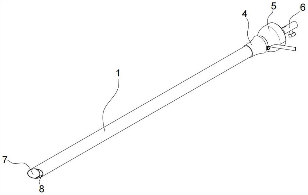
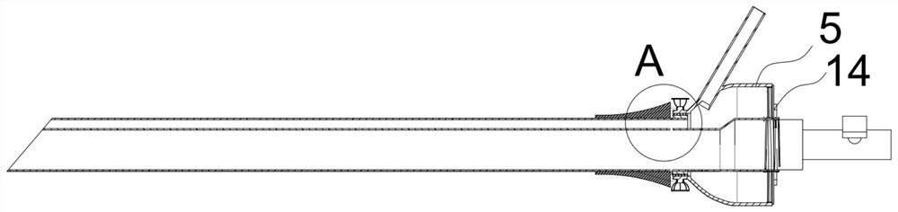
[0088] Such as Figure 1~5 As shown, the present invention discloses a percutaneous renal ultramicrochannel visual puncture and dilation integrated kit with negative pressure suction, including:

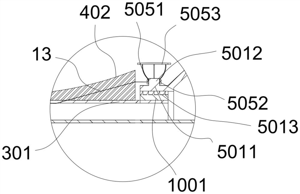
[0089] A sheath 1 having an inner open end at a distal end 101 of the sheath, and an outer open end at a proximal end 102 of the sheath, which is used for introduction into a body requiring a surgical operation In the region, the sheath tube 1 is provided with a negative pressure channel 7 and a perfusion channel 8 that are independent of each other, and the negative pressure channel 7 and the perfusion channel 8 are separated by the partition wall 302;

[0090] The expansion tube joint 5 is connected to the outer opening end of the sheath tube proximal end 102, and a catheter device 504 is arranged on the expansion tube joint 5, and the catheter device 504 can inject irrigation water into the perfusion channel 8 and / or operate instruments ;

[0091] The negative pressure output de...


PUM


Abstract

The invention provides a percutaneous kidney ultramicro-channel visible puncture and expansion integrated kit with negative pressure suction. The percutaneous kidney ultramicro-channel visible puncture and expansion integrated kit comprises a sheath tube, an expansion tube connector and a negative pressure output device, the sheath tube is provided with an inner opening end at the far end of the sheath tube and an outer opening end at the near end of the sheath tube, a negative pressure channel and a perfusion channel which are independent of each other are arranged in the sheath tube, and the negative pressure channel and the perfusion channel are separated by a partition wall; the expansion tube connector is connected with the outer opening end of the near end of the sheath tube, a catheter device is arranged on the expansion tube connector, and the catheter device can inject flushing water into the perfusion channel or operate instruments; and the negative pressure output device is connected with the outer opening end at the near end of the sheath tube, and the negative pressure output device is hermetically communicated with the negative pressure channel. By improving the structure of the sheath tube, the sheath tube meets the functional requirements of liquid injection and stone suction, wide visual field and clear images are guaranteed, meanwhile, large stone fragments can be sucked and discharged, repeated work is avoided, the operation time is shortened, the operation efficiency is improved, and the work intensity of doctors is reduced.

Description

technical field [0001] The invention relates to the technical field of medical aids, in particular to a percutaneous kidney ultramicro channel visual puncture and expansion integrated set with negative pressure suction. Background technique [0002] Kidney stones are one of the most common diseases in urology. Percutaneous nephroscopic surgery is a minimally invasive technique widely used in the treatment of kidney stones. Surgical method for lithotripsy. [0003] In the past 20 years, scholars at home and abroad have made unremitting efforts for the minimally invasive percutaneous renal passage, and designed a series of minimally invasive PCNL, including minimally invasive percutaneous nephrolithotomy (mini-mally invasive PCNL). , MPCNL), Chinese MPCNL (Chinese MPCNL), ultra-fine percutaneous nephrolithotomy (ultra-mi ni-PCNL, UMP), micro-channel percutaneous nephrolithotripsy (Micro-PCNL) and so on. In practice, people have gradually realized that there are problems such...

Claims


Application Information

Patent Timeline
Patent Type & Authority Applications(China)
IPC IPC(8): A61B18/26A61B90/00A61B17/34
CPCA61B18/26A61B90/08A61B17/3403A61B2090/08021A61B2217/005A61B2217/007A61B2218/002A61B2218/007A61B2017/3413
Inventor 胡嘉盛
Owner NINGBO FIRST HOSPITAL
Why Eureka
Try Eureka