
Pathological hard tissue slicing system

A hard tissue, pathological technology, applied in the field of pathological experiments, can solve the problems of pathologist's operation error, instability, and increased scrapping rate of diseased tissue.

Pending Publication Date: 2021-12-28
澎立检测技术(上海)有限公司
View PDF0 Cites 1 Cited by
  • Summary
  • Abstract
  • Description
  • Claims
  • Application Information

AI Technical Summary

Problems solved by technology

[0004] Now it is commonly used to manually operate and identify the above steps through pathologists. However, due to the large gap of pathologists, the pathologists in each hospital are very busy with work, and there is a lot of backlog work. Block cutting is unstable, and pathologists will also make operational errors, which will lead to an increase in the scrap rate of diseased tissues and an increase in procedures for meaningless operations

Method used


Image

  • Pathological hard tissue slicing system
  • Pathological hard tissue slicing system
  • Pathological hard tissue slicing system

Examples


specific Embodiment 1

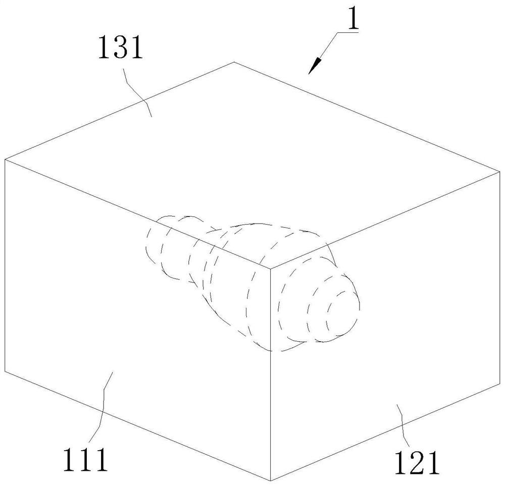
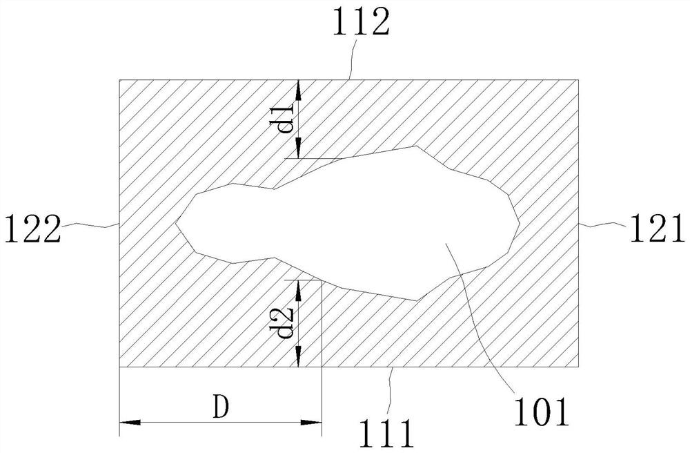
[0050] Specific embodiment one: please refer to Figure 1-8 A pathological hard tissue slicing system, used for slicing a wax block 1 embedded with pathological tissue 101, the wax block 1 is a light-transmitting cuboid or cube. The wax block 1 includes a left side 111 and a right side 112 which are mutually symmetrical planes, a front side 121 and a rear side 122 which are mutually symmetrical planes, an upper side 131 and a lower side 132 which are mutually symmetrical planes. The slicing system includes a distance-fixing unit 2, a clamping unit 3, a monitoring camera 4, a cutting unit 5 and a control unit.

[0051] The distance-fixing unit 2, the clamping unit 3, the monitoring camera 4 and the cutting unit 5 are all controlled by the control unit.

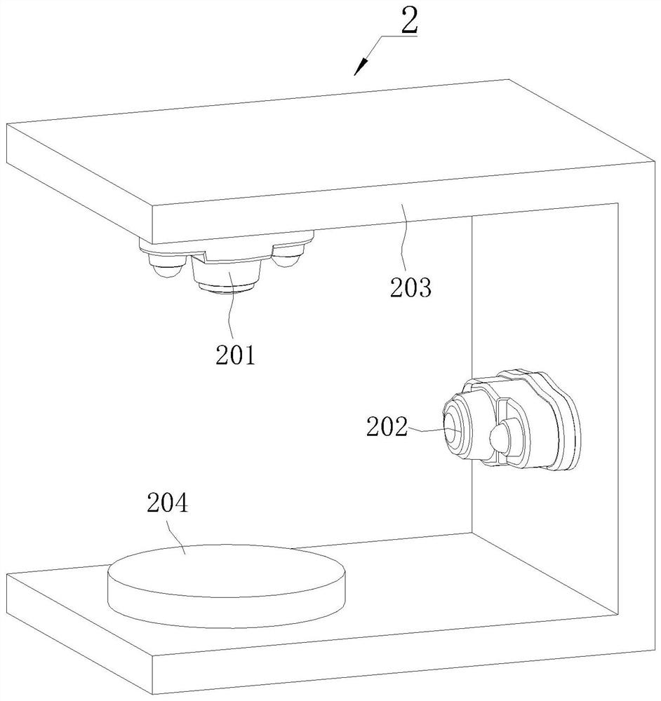
[0052] The distance-fixing unit 2 collects distance information on the wax block 1 through the upper camera 201 and the side camera 202 to determine the maximum section position of the pathological tissue 101 .

[0053] The d...

specific Embodiment 2

[0083] Embodiment 2: Different from Embodiment 1, the cutting unit 5 includes a blade 501 and a cutting motor 502 . The output end of the cutting motor 502 is connected with the blade 501, and the output end of the cutting motor 502 can move vertically up and down, so that the blade 501 can cut part of the wax block 1 located outside the cutting end face. After the blade 501 completes a cycle of moving up and down, the movable top block 305 moves to the outside by a set distance, and the set distance is set by the control unit. Effectively reduce the setting of electrical components and reduce costs.

specific Embodiment 3

[0084] Embodiment 3: On the basis of Embodiment 1 or 2, the clamping unit 3 further includes writing brushes and crayons.

[0085] The writing brush is driven by the writing brush motor, and the writing brush motor is controlled by the control unit.

[0086] The crayon is driven by a crayon motor, which is controlled by a control unit.

[0087] During rough cutting, after the blade 501 completes a cycle of moving up and down, the control unit controls the operation of the brush motor to make the brush sweep the cut surface of the wax block 1, so that the paraffin burrs produced after rough cutting are swept away.

[0088] When finely cutting, after the blade 501 completes a cycle of moving up and down, the control unit controls the operation of the crayon motor, so that the crayon holds up the cut slices and puts them into the pool, waiting for the next step of dyeing process.

[0089] It effectively reduces the manual work and reduces the problem of chip rupture caused by th...


PUM


Abstract

The invention discloses a pathological hard tissue slicing system, and belongs to the field of pathological experiments, the pathological hard tissue slicing system is used for slicing a wax block embedded with pathological tissue, the pathological hard tissue slicing system comprises a distance fixing unit, a clamping unit, a monitoring camera, a cutting unit and a control unit; the distance fixing unit collects distance information of the wax block through a visual element; the clamping unit is used for clamping and fastening a wax block, the clamping unit can control the clamping length of the wax block, the cutting unit comprises a blade, the monitoring camera is used for collecting structural information of the wax block, and the distance fixing unit, the clamping unit, the monitoring camera and the cutting unit are all controlled by the control unit. The position of the maximum section of the diseased tissue can be pre-judged; the wax block is roughly cut to the maximum section of the diseased tissue rapidly and accurately, then precise slice cutting is conducted, meanwhile, factors of the voidage, the tissue continuity and the tissue hierarchical sequence of the wax block are monitored in real time, unqualified wax blocks are rapidly removed, invalid actions are reduced, and the cutting efficiency is improved.

Description

technical field [0001] The invention belongs to the field of pathological experiments, and more specifically relates to a pathological hard tissue slicing system. Background technique [0002] In medical diagnosis, it is usually necessary to take a pathology of a predetermined size from a diseased part of a patient to make a pathological section. Pathological section refers to taking a certain size of diseased tissue and making pathological sections with histopathological methods. Usually, the diseased tissue is embedded in paraffin block, cut into thin slices with a microtome, and then stained with a microscope. Examine the lesion, the occurrence and development process of the lesion, and finally make a pathological diagnosis. [0003] When performing pathological tissue slicing, it is necessary to freeze and harden the wax block embedded with diseased tissue first, and then expose the largest section of the diseased tissue with the cutting knife of the wax block by rough ...

Claims


Application Information

Patent Timeline
Patent Type & Authority Applications(China)
IPC IPC(8): G01N1/06G01N1/28B26D7/02B26D5/00B26D1/06
CPCG01N1/06G01N1/28B26D1/06B26D5/007B26D7/025
Inventor 袁红江吕见
Owner 澎立检测技术(上海)有限公司
Learn More