Intraoperative ultrasonic probe sleeve and use method thereof

An ultrasonic probe and probe cover technology, which is applied in ultrasonic/sonic/infrasonic diagnosis, application, and sonic diagnosis, etc., can solve the problems of unclear ultrasonic image display of couplant, cumbersome socketing procedures, affecting operation time, etc. The connection procedure is cumbersome, the use and operation are convenient, and the effect of avoiding spillover and waste

Pending Publication Date: 2021-12-31
四川省妇幼保健院
View PDF0 Cites 0 Cited by
  • Summary
  • Abstract
  • Description
  • Claims
  • Application Information

AI Technical Summary

Problems solved by technology

[0005] The purpose of the present invention is to provide an intraoperative ultrasonic probe cover and its use method, so as to solve the problem of cumbersome plastic sleeve fitting procedures in the prior art, unsatisfactory effect, affecting operation time, and unclear display of ultrasonic images caused by coupling agent overflow. technical problem

Method used


Image

  • Intraoperative ultrasonic probe sleeve and use method thereof
  • Intraoperative ultrasonic probe sleeve and use method thereof

Examples


Embodiment 1

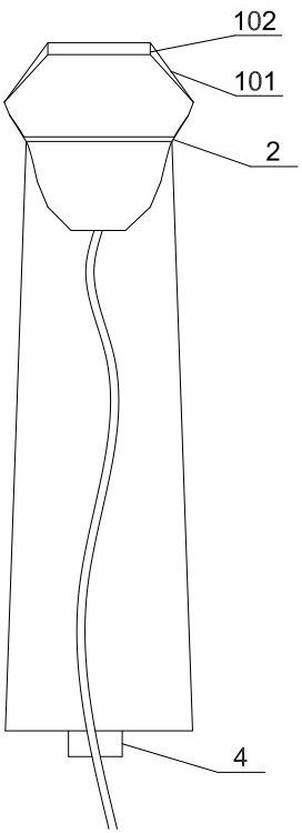
[0029] Such as Figure 1-2 Shown:

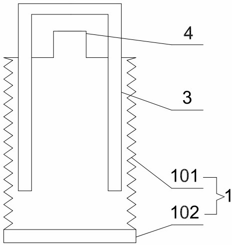
[0030] The present invention provides an intraoperative ultrasonic probe cover, which includes a cover body 1, a fastening ring 2 for fixing the cover body 1 to the ultrasonic probe, and a shaping block 3 for supporting the cover body 1;

[0031] The cover body 1 includes a probe cover film 101 capable of wrapping an ultrasonic probe and an ultrasonic probe line and a coupling patch 102 arranged inside the center of the probe cover film 101. The coupling patch 102 matches the size of the ultrasonic probe. The cover film 101 matches the size of the ultrasonic probe and can fit closely;

[0032] When the cover body 1 is stored for use, the coupling patch 102 is set outward, and the probe cover film 101 is folded to form a hollow structure, and the size of the shaped block 3 matches the inner diameter of the probe cover film 101, and is supported by the probe cover film 101 for folding formed hollow structure.

[0033] As an optional embodim...

Embodiment 2

[0039] Compared with Embodiment 1, the present invention provides an intraoperative ultrasound probe cover, which also has the following differences:

[0040] The shaped block 3 is an "H" shaped structure.

[0041] As an optional embodiment, the coupling patch 102 is a solid hydrogel material with a wet surface, flexibility and ductility, and a thickness of 3 mm.

[0042] As an optional implementation, the probe cover film 101 is made of plastic.

[0043] As an optional implementation manner, the length of the probe cover film 101 is 80 cm.

Embodiment 3

[0045] Compared with Embodiment 1, the present invention provides an intraoperative ultrasound probe cover, which also has the following differences:

[0046] Described stereotyped block 3 is " N " type structure.

[0047] As an optional embodiment, the coupling patch 102 is a solid hydrogel material with a wet surface, flexibility and ductility, and a thickness of 1.5 mm.

[0048] As an optional implementation, the probe cover film 101 is made of latex.

[0049] As an optional implementation manner, the length of the probe cover film 101 is 100 cm.


PUM


Abstract

The invention discloses an intraoperative ultrasonic probe sleeve and a use method thereof, and solves the technical problems that in the prior art, an endoscope plastic sleeve is tedious in sleeving procedure and not ideal in effect, the operation time is affected, a coupling agent overflows, and an ultrasonic image is not clearly displayed. The intraoperative ultrasonic probe sleeve comprises a sleeve body (1), a fastening ring (2) for fixing the sleeve body (1) on an ultrasonic probe, and a shaping block (3) for supporting the sleeve body (1); and the sleeve body (1) comprises a probe sleeve film (101) capable of wrapping the ultrasonic probe and an ultrasonic probe line and a coupling patch (102) arranged on the inner side of the center of the probe sleeve film (101). According to the intraoperative ultrasonic probe sleeve, the defects of tedious sleeving procedure, non-ideal effect and influence on operation of the existing intraoperative endoscope plastic sleeve are avoided, the probe sleeve sleeving efficiency and the probe moving flexibility during examination by a doctor are greatly improved, the time cost and the labor cost of the doctor are saved, and the working efficiency and the working quality of the doctor are improved.

Description

technical field [0001] The invention relates to a protective cover for medical instruments, in particular to an intraoperative ultrasonic probe cover and a use method thereof. Background technique [0002] When doctors perform minimally invasive breast surgery, or when doctors need intraoperative ultrasound in open surgery, they often need ultrasound-guided clinical operations. At this time, the ultrasound probe needs to be in a sterile environment. For this, clinical work often uses The method is to apply coupling agent on the surface of the probe, then use the plastic sleeve of the sterile endoscope to connect the probe and the probe wire, and then use gauze strips to ligate the concave part of the probe handle to fix it. This method is cumbersome, time-consuming and laborious, and seriously increases the The cost of time and labor for doctors, and the effect is often not ideal, the gauze strips are often not tightly ligated, and the ultrasonic coupling agent in the plasti...

Claims


Application Information

Patent Timeline
Patent Type & Authority Applications(China)
IPC IPC(8): A61B8/00
CPCA61B8/4422A61B8/4444A61B8/4281
Inventor 梁蓉赵婧何冠南白艳杨家翔罗静闫梅李宁
Owner 四川省妇幼保健院
Why Eureka
Try Eureka