
Lung disease lesion unsupervised segmentation method based on knowledge distillation

A lung disease, unsupervised technology, applied in the fields of medical image processing and computer vision, can solve problems such as insufficient performance and insufficient knowledge distillation, and achieve the effects of easy collection, easy construction, and insufficient labeled data

Pending Publication Date: 2022-01-07
DALIAN UNIV OF TECH
View PDF0 Cites 5 Cited by
  • Summary
  • Abstract
  • Description
  • Claims
  • Application Information

AI Technical Summary

Problems solved by technology

But directly applying the framework to CT images does not give full play to its capabilities, and its teacher network is usually pre-trained on natural image datasets, which are quite different from CT image datasets.
In addition, distillation only at the pixel level ignores the relationship between pixels, and knowledge distillation is not sufficient

Method used


Image

  • Lung disease lesion unsupervised segmentation method based on knowledge distillation
  • Lung disease lesion unsupervised segmentation method based on knowledge distillation
  • Lung disease lesion unsupervised segmentation method based on knowledge distillation

Examples


Embodiment Construction

[0052] The unsupervised segmentation method of lung disease lesions based on knowledge distillation proposed by the present invention will be described in detail below in conjunction with examples and accompanying drawings:

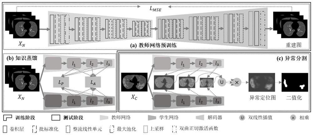
[0053] The present invention designs pixel-level and affinity-level knowledge distillation to realize unsupervised segmentation of lung disease lesions, and only uses normal CT images for training to achieve more accurate lesion segmentation. The specific implementation process is as figure 1 As shown, the method includes the following steps:

[0054] 1) Collect initial data:

[0055] 1-1) Collect the training data set. The experiment uses a private normal data set. A total of 69 normal lung CT images are collected for training, and the axial image resolution is 512×512.

[0056] 1-2) Collect test data sets. The experiment uses three new coronary pneumonia data sets for testing, all of which are lung CT images diagnosed with new coronary pneumonia. Dat...


PUM


Abstract

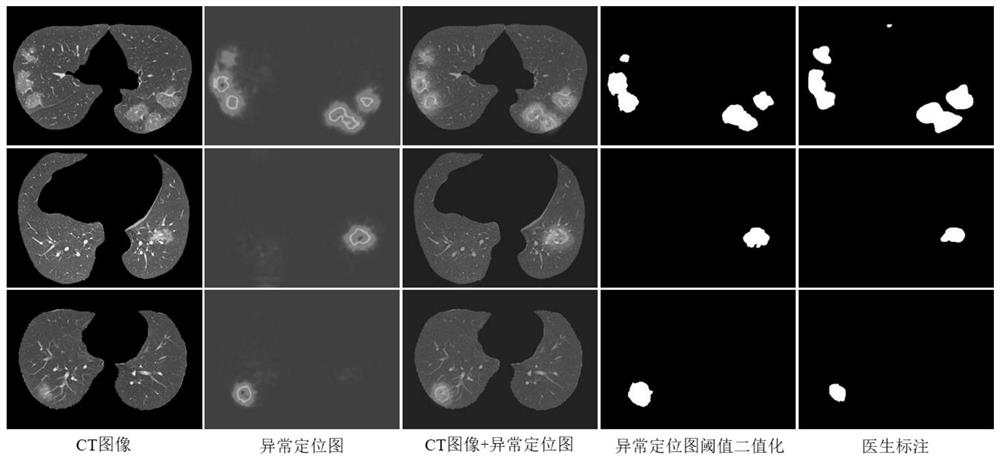
The invention discloses a lung disease lesion unsupervised segmentation method based on knowledge distillation, and belongs to the field of medical image processing and computer vision. The method comprises the following steps: firstly, constructing and training an auto-encoder to obtain a pre-trained teacher network with rich CT image semantic knowledge, then training a student network with the same architecture as the teacher network from the pre-trained teacher network by only distilling the knowledge of normal CT images, and finally, performing lesion segmentation on the difference of features extracted by the lesion-containing image by using teacher and student networks. Meanwhile, besides traditional pixel-level distillation, affinity-level distillation considering the relation between pixels is further designed in the method, so that effective knowledge is fully distilled. Experiments prove that the lesion segmentation precision can be effectively improved on different data sets. The method is easy to construct, an unlabeled lung disease focus segmentation result can be obtained only depending on normal data, and generalization and operation efficiency are considerable.

Description

technical field [0001] The invention belongs to the field of medical image processing and computer vision, and relates to pixel-level segmentation of lung disease lesions in computer tomography (Computed Tomography, CT) images by using a deep learning neural network framework, in particular to knowledge distillation-based lung A method for unsupervised segmentation of disease lesions. Background technique [0002] In recent years, the impact of lung diseases on people's health has become increasingly serious, and the computer-aided diagnosis (Computer Aided Diagnosis, CAD) system based on chest CT images is very important for the rapid diagnosis and evaluation of diseases. Usually, such CAD systems include an important processing step, which is automatic segmentation of lesions on CT images. Most of the existing work uses supervised methods for lesion segmentation (Huang L, Han R, Ai T, et al. Serial quantitative chest CTassessment of COVID-19: Deep-learning approach [J]. R...

Claims


Application Information

Patent Timeline
IPC IPC(8): G06T7/11G06N5/02
CPCG06T7/11G06N5/02G06T2207/10081G06T2207/20081G06T2207/20221G06T2207/30061G06T2207/30096
Inventor 徐睿王宇凤叶昕辰
Owner DALIAN UNIV OF TECH
Learn More