Radiotherapy dose verification method

A dose verification and dose technology, applied in radiation therapy, X-ray/γ-ray/particle irradiation therapy, treatment, etc., can solve the problems of different density values ​​and phantoms of EPID equipment, and achieve high-precision results

Active Publication Date: 2022-03-04
BEIJING INSTITUTE OF TECHNOLOGYGY
View PDF18 Cites 0 Cited by
  • Summary
  • Abstract
  • Description
  • Claims
  • Application Information

AI Technical Summary

Problems solved by technology

This method can generally obtain a relatively satisfactory three-dimensional radiation dose in the phantom, but there are still some defects in this method, mainly: ①The dose conversion of this method is realized on the EPID plane, and the gray value of the EPID plane Converted to a dose value, then back-calculated into the phantom by exponential decay
However, the dose value released by X-rays is related to the mass-energy attenuation coefficient, and the density value of the EPID device is different from the phantom, so the relationship between the two cannot be converted by simple exponential attenuation

Method used


Image

  • Radiotherapy dose verification method
  • Radiotherapy dose verification method
  • Radiotherapy dose verification method

Examples


Embodiment

[0083] The accelerator used in this embodiment is Varian C2300, equipped with HD120 multi-leaf grating, which can release X-rays with a ray energy of 6MV for radiation therapy; the EPID model used is aS1000, with an effective detection area of ​​30cm×40cm, and the imaging size 768×1024 pixels, the size of a single pixel is about 0.039cm×0.039cm, the distance between the upper surface of the EPID and the accelerator source is 150cm, background correction and pan-field correction have been performed on the EPID before image acquisition. The acquisition software uses Varian's IAS3 (Image Acquistion System3), the acquired EPID images are exported through Aria software, and the subsequent dose verification is realized on the MATLAB2019a platform.

[0084] The parameter modeling of the radiotherapy equipment is realized by using the solid water phantom, and then the dose verification is carried out on the CIRS simulation chest model under the same X-ray irradiation conditions, such a...


PUM


Abstract

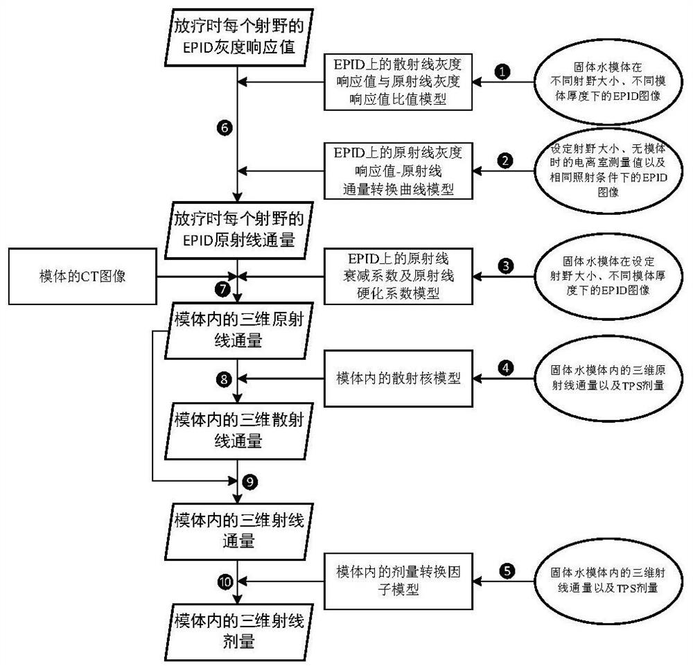
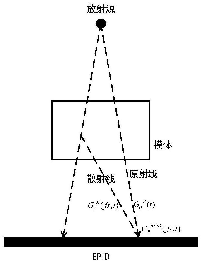
The invention relates to a radiotherapy dose verification method, and belongs to the technical field of dose verification in a radiotherapy process. The method comprises the following steps of: firstly, extracting an original ray gray scale response value on the EPID from a gray scale response value on the EPID, converting the original ray gray scale response value into an original ray flux on the EPID, then reversely calculating an original ray flux value of each point in a die body, then determining a scattered ray flux value in the die body by using a scattering kernel in the die body, and finally, calculating an original ray flux value of each point in the die body. Using the dose conversion factor in the die body to obtain a dose value of a corresponding position; in the prior art, dose conversion is achieved on EPID in a fitting mode, and due to the fact that actual dose absorption of a certain position in the body of a patient in the radiotherapy process is related to the flux of X (gamma) rays penetrating through the position and is not directly related to the flux on the EPID, compared with the prior art, dose conversion factors of the method better conform to the actual physical process, and the dose conversion efficiency is greatly improved. And the precision of the reconstructed dose is higher.

Description

technical field [0001] The present invention relates to a radiotherapy dose verification method. The method calculates the three-dimensional dose through direct inversion based on EPID (electronic portal image device), and then compares the three-dimensional radiation dose in the phantom with the TPS dose to realize radiotherapy dose verification. The invention belongs to the technical field of dose verification during radiotherapy. Background technique [0002] Existing radiotherapy dose verification methods can be divided into two categories: forward method and reverse method. The main disadvantage of the forward method is that the dose difference can only be compared on the two-dimensional plane, and the relationship between the dose difference on the EPID plane and the dose difference in the patient is not yet clear. In order to achieve precise radiotherapy, the reverse method must be used. The latest development of the reverse method was proposed by M.Wending et al. i...

Claims


Application Information

Patent Timeline
IPC IPC(8): A61N5/10
CPCA61N5/103A61N5/1031A61N5/1049A61N5/1075A61N2005/1054A61N2005/1076
Inventor 温俊海张军李杰
Owner BEIJING INSTITUTE OF TECHNOLOGYGY
Why Eureka
Try Eureka