Preparation method of 3D printing stent for maintaining specimen skin branch chain shape

A 3D printing and specimen technology, applied in the field of digital human anatomy teaching, can solve problems such as morphological changes, difficulty in meeting the high quality requirements of perforator and cutaneous branch angiography images, and artifact interference.

Pending Publication Date: 2022-03-29
广州医科大学附属顺德医院
View PDF14 Cites 0 Cited by
  • Summary
  • Abstract
  • Description
  • Claims
  • Application Information

AI Technical Summary

Problems solved by technology

However, various artifact reduction methods are still difficult to meet the high-quality requirements of perforator and cutaneous branch angiographic images.
[0003] To sum up, the current digital angiography technology of perforator and cutaneous branch vessels often has problems such as artifact interference and morphological changes. If we want to accurately analyze the anastomotic characteristics of perforator and cutaneous branch vessels, we need to solve these problems

Method used


Image

  • Preparation method of 3D printing stent for maintaining specimen skin branch chain shape
  • Preparation method of 3D printing stent for maintaining specimen skin branch chain shape
  • Preparation method of 3D printing stent for maintaining specimen skin branch chain shape

Examples


Embodiment Construction

[0025] The following is a further detailed description through specific embodiments:

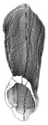
[0026] A preparation method of a 3D printed scaffold for maintaining the branched-chain morphology of a specimen skin, comprising the following steps:

[0027] S1. Perform the first spiral CT scan after perfusion of the fresh specimen to obtain data set 1;

[0028] S2. After peeling off the skin and superficial fascia of the specimen, perform a second spiral CT scan to obtain data set 2;

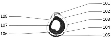
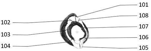
[0029] S3. Use MIMICS software to carry out 3D reconstruction of the continuous 2D tomographic images of dataset 2, and select the overall mask after 3D reconstruction as mask 1;

[0030] S4. After batch scaling the 2D tomographic images, select the overall mask for 3D reconstruction and save it as mask 2;

[0031] S5. After registering the center line of the long axis of the mask 1 and the mask 2, remove the overlapping part to obtain the hollow mask 3;

[0032] S6. Divide the mask 3 according to the siz...


PUM

PropertyMeasurementUnit
diameteraaaaaaaaaa

Abstract

The invention discloses a preparation method of a 3D printing stent for maintaining the form of a skin branch chain of a specimen, which comprises the following steps of: scanning the specimen for multiple times to obtain a plurality of data sets; the data sets are processed through MIMICS software, and masks are obtained; the mask with the parting lines is imported into 3D printing software; 3D printing equipment is used for printing the support. According to the method, based on the digital dissection and 3D printing technology, the skin scaffold completely matched with the specimen soft tissue shape is manufactured, the original shape of the specimen skin is supported and maintained, and high-density tissue and heterogeneous material artifacts are avoided; according to the method, a fresh specimen filled with red lead oxide latex can be separated in order, and the cut skin and superficial fascia are supported and subjected to microCT scanning, so that surgeons and dissection researchers can analyze skin branch contrast scanning data and fuse images in order; and for a large-range skin flap penetrating branch and skin branch chain structure, the difference and relation between skin branch chains at different anatomical parts can be more comprehensively understood.

Description

technical field [0001] The invention relates to the field of digital human anatomy teaching, in particular to a preparation method of a 3D printing bracket for maintaining the branched chain shape of the specimen skin. Background technique [0002] The selection and application of skin flaps is a common surgical technique in reconstructive plastic surgery. The damage of the donor site is small, the function of the recipient site is recovered well after operation, and the appearance is beautiful. Although the surgical technique of skin flap extraction is becoming more and more perfect, with the development of 3D visualization digital anatomy technology and minimally invasive surgery, surgeons and Anatomical researchers have increasingly stringent requirements for flap design. They hope to present the vascular shape of the flap more intuitively, analyze the perforator region and the distribution of blood vessels more accurately, and help students better understand the perforat...

Claims


Application Information

Patent Timeline
Patent Type & Authority Applications(China)
IPC IPC(8): B29C64/10B29C64/386G09B23/30G09B23/28B33Y10/00B33Y50/00
CPCB29C64/10B29C64/386G09B23/30G09B23/286B33Y10/00B33Y50/00
Inventor 何藻鹏秦向征张琳李卫
Owner 广州医科大学附属顺德医院
Why Eureka
Try Eureka