Eureka AIR delivers breakthrough ideas for toughest innovation challenges, trusted by R&D personnel around the world.

Angiocardiography device

A cardiovascular and coaxial technology is applied in the field of cardiovascular angiography devices to achieve the effects of convenient angiography operation, simple and convenient operation, and convenient operation.

Pending Publication Date: 2022-04-05
SHANDONG FIRST MEDICAL UNIV & SHANDONG ACADEMY OF MEDICAL SCI
View PDF0 Cites 2 Cited by
  • Summary
  • Abstract
  • Description
  • Claims
  • Application Information

AI Technical Summary

Problems solved by technology

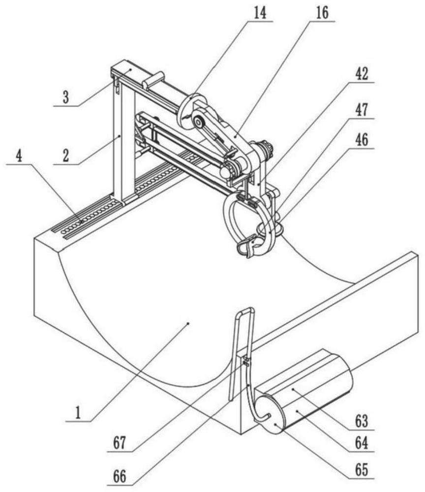
[0003] Aiming at the deficiencies of the prior art, the present invention provides a cardiovascular angiography device, which can clamp and fix the puncture needle and dilation tube, and can help the doctor to compress the medical gauze and assist the doctor to perform the angiography operation, effectively solving the above background problems mentioned in the technology

Method used


Image

  • Angiocardiography device
  • Angiocardiography device
  • Angiocardiography device

Examples


Embodiment Construction

[0035] The following are specific embodiments of the present invention, and further describe the technical solution of the present invention in conjunction with the accompanying drawings, but the present invention is not limited to these embodiments.

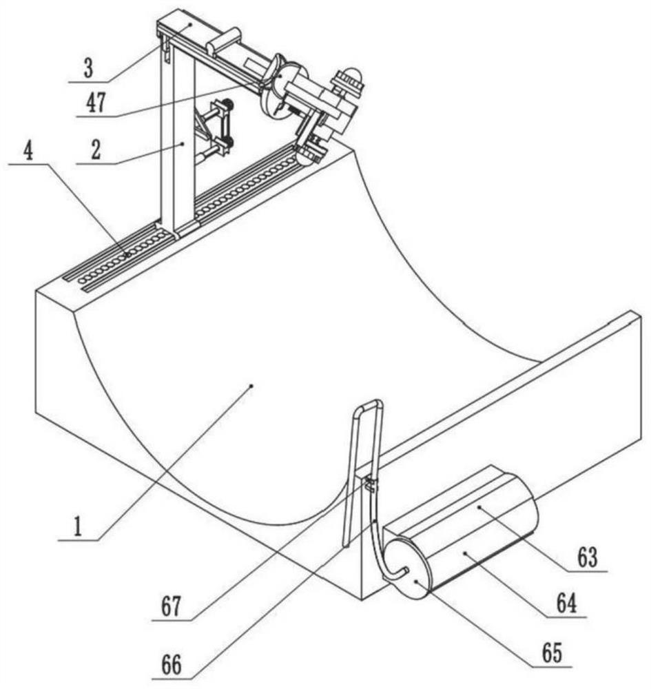
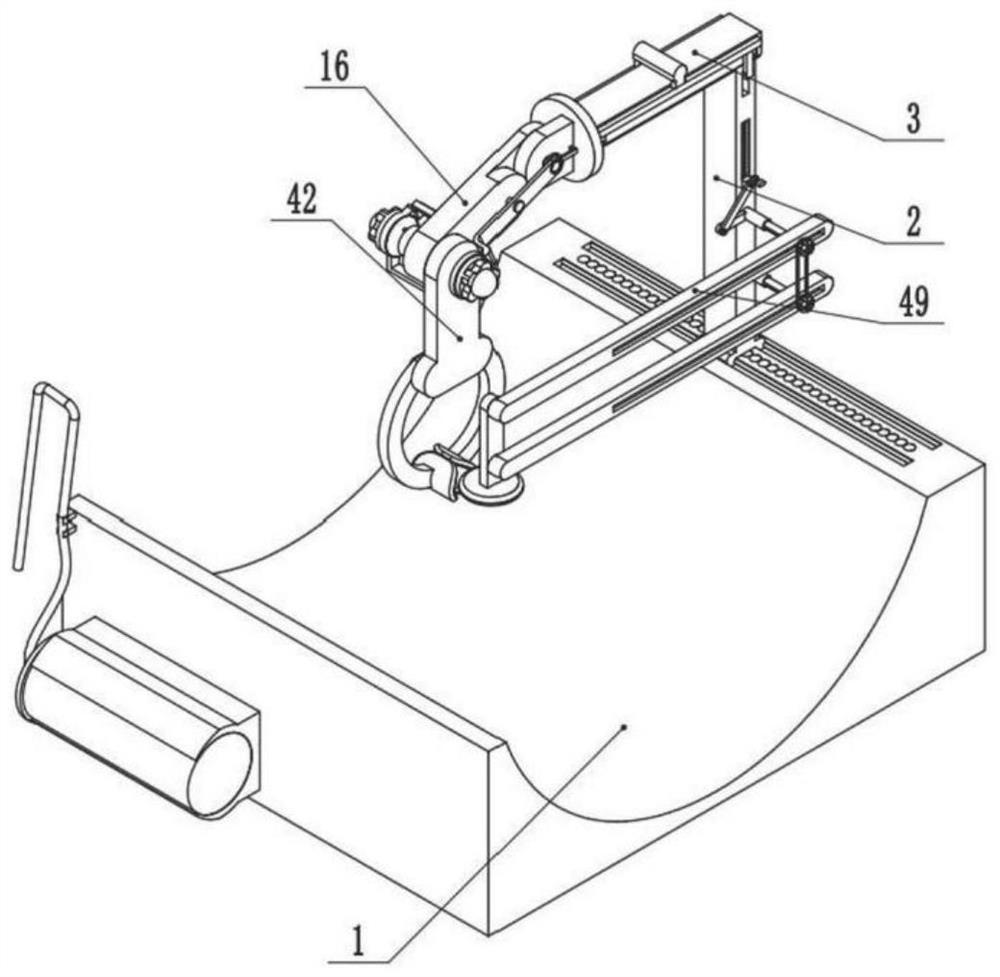
[0036] Such as Figure 1-19 As shown, the present invention provides a cardiovascular imaging device, including a support pad 1, the upper end of the support pad 1 is slidably connected with a support column 2 that can move left and right, and the upper end of the support column 2 is slidably connected with a support column that can move forward and backward. Moving adjustment plate 3, the upper end rear side of the support pad 1 is provided with a plurality of first positioning holes 4, the lower end of the adjustment plate 3 is provided with a plurality of second positioning holes 5, and the upper and lower sides of the support column 2 are The two sides are respectively slidably connected with a first positioning column 6 and...


PUM


Abstract

An angiocardiography device is characterized in that a supporting column capable of moving left and right is slidably connected to the rear side of the upper end of a supporting pad, an adjusting plate capable of moving front and back is slidably connected to the upper end of the supporting column, a butt-joint plate is rotatably connected to the front end of the adjusting plate, and a first adjusting arm capable of swinging up and down is hinged to the front end of the butt-joint plate; a second adjusting arm capable of swinging up and down is hinged to the front end of the first adjusting arm, clamping arms capable of synchronously swinging outwards are hinged to the front side and the rear side of the lower side of the left end of the second adjusting arm respectively, and clamping arc plates are fixedly connected to the inner sides of the lower ends of the clamping arms respectively; when the device is used, the clamping arc plate can be effectively and accurately adjusted according to the position and angle of a puncture needle or an expansion tube, the puncture needle or the expansion tube can be conveniently and accurately fixed through the clamping arc plate, operation of a doctor is greatly facilitated, operation is easy and convenient, and radiography operation of the doctor is greatly facilitated.

Description

technical field [0001] The invention relates to the field of medical equipment, in particular to a cardiovascular imaging device. Background technique [0002] Cardiovascular angiography is to quickly inject contrast agent into the heart cavity or blood vessel through the cardiac catheter, so that the heart and blood vessel cavity can be visualized under X-ray irradiation. After the development process is photographed, the blood flow sequence containing the contrast agent and the filling of the heart vessels can be seen from the results of the development, so as to understand the physiological and anatomical changes of the heart and blood vessels; when performing cardiovascular angiography, it is first necessary to puncture the human body After the puncture is successful, a dilation tube is inserted, and then the guide wire is inserted deep into the heart cavity, and then the catheter is inserted into the heart cavity along the guide wire. After the guide wire is pulled out,...

Claims


Application Information

Patent Timeline
IPC IPC(8): A61B17/34A61B90/14A61B6/00
Inventor 宋晓瑞王学政赵晓辉邱建峰路伟钊马骏驰侯坤王宝剑冯子洋
Owner SHANDONG FIRST MEDICAL UNIV & SHANDONG ACADEMY OF MEDICAL SCI
Why Eureka
Eureka Blog
Learn More