Method for automatically detecting and displaying ultrasonic craniocerebral abnormal region

An abnormal area, automatic detection technology, applied in image data processing, instruments, calculations, etc., can solve the problems of complex brain ultrasound images, and achieve the effect of improving the accuracy.

Pending Publication Date: 2022-04-26
SHANTOU INST OF UITRASONIC INSTR CO LTD
View PDF0 Cites 2 Cited by
  • Summary
  • Abstract
  • Description
  • Claims
  • Application Information

AI Technical Summary

Problems solved by technology

However, the reason why traditional processing and popular machine learning methods cannot be effectively applied to cranial ultrasound image analysis and diagnosis lies in the complexity of cranial ultrasound image itself.

Method used


Image

  • Method for automatically detecting and displaying ultrasonic craniocerebral abnormal region

Examples


Embodiment 1

[0017] Embodiment 1, a method for automatic detection and display of abnormal brain regions by ultrasound, comprising the following steps:

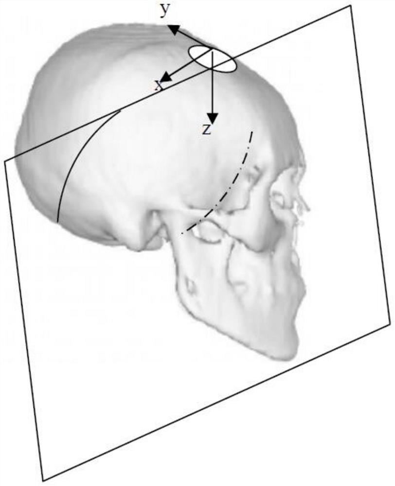
[0018] S01. Construct the curved surface model of the cranium first, and construct the curved surface model of the cranium according to the ultrasonic images obtained by the ultrasonic scanning of the cranium.

[0019] S02. Perform cranial edge detection on the 2D ultrasonic image obtained by ultrasonic scanning of the cranium to obtain a cranial edge curve of the 2D image.

[0020] S03. Fitting the cranial edge curve of the 2D image obtained in step S02 with the cranial surface model obtained in step S01 to determine the position of the 2D image on the cranial surface model.

[0021] S04. According to the position of the 2D image obtained in step S03 on the curved skull model, it is judged whether the 2D image is symmetrical with respect to the midsagittal plane or the median coronal plane of the curved skull model.

[0022] S05. Mark t...

Embodiment 2

[0027] Embodiment 2, a method for automatic detection and display of ultrasonic brain abnormalities, comprising the following steps:

[0028] S01. Construct the curved surface model of the cranium first, and construct the curved surface model of the cranium according to the ultrasonic images obtained by ultrasonic scanning of the cranium.

[0029] S02. Perform cranial edge detection on the 2D ultrasonic image obtained by ultrasonic scanning of the cranium to obtain a cranial edge curve of the 2D image.

[0030] S03. Fitting the cranial edge curve of the 2D image obtained in step S02 with the cranial surface model obtained in step S01 to determine the position of the 2D image on the cranial surface model.

[0031] S04. According to the position of the 2D image obtained in step S03 on the curved skull model, it is judged whether the 2D image is symmetrical with respect to the midsagittal plane or the median coronal plane of the curved skull model.

[0032] S05. Mark the 2D imag...


PUM


Abstract

The invention relates to the field of ultrasonic detection, in particular to an automatic detection and display method for an ultrasonic craniocerebral abnormal area. According to the technical scheme, firstly, a skull curved surface model is constructed, then edge detection is conducted on a 2D ultrasonic image to obtain a skull edge curve, and the edge curve and the skull curved surface model are used for fitting to determine the position of the 2D ultrasonic image so as to judge whether the 2D image has the symmetry characteristic or not; and finally, carrying out similarity comparison calculation on the two symmetrical regions by utilizing the symmetry characteristic of the 2D image so as to determine whether an abnormal region exists and determine the position of the abnormal region. The method has the advantages that the skull curved surface model is firstly established, the skull boundary curve of the 2D ultrasonic image is detected, the skull boundary curve and the skull curved surface model are fitted to determine the specific position of the 2D image so as to select the 2D image with the symmetry characteristic, and the abnormal area is detected, segmented and displayed by using the symmetry of the 2D image. Therefore, the accuracy of abnormal region detection is effectively improved.

Description

technical field [0001] The invention relates to the field of ultrasonic detection, in particular to a method for automatic detection and display of ultrasonic brain abnormalities. Background technique [0002] Ultrasound brain scan is a routine clinical practice, especially in the clinical monitoring of newborns is widely used. Ultrasound cranial scan is mainly used for the diagnosis of intraventricular hemorrhage, pericerebral hemorrhage infarction, ventriculomegaly after hemorrhage, and fluid pericranial leukomalacia / paralysis after hemorrhage. In traditional diagnosis, ultrasound doctors rely on knowledge and experience to identify abnormalities in images; with the continuous development of computer technology, using computers to automatically analyze and diagnose images can reduce the burden on doctors' diagnosis and improve efficiency. However, the reason why traditional processing and popular machine learning methods cannot be effectively applied to cranial ultrasound...

Claims


Application Information

Patent Timeline
Patent Type & Authority Applications(China)
IPC IPC(8): G06T7/00G06T7/13G06T7/174G06T7/68
CPCG06T7/0014G06T7/13G06T7/174G06T7/68G06T2207/10132G06T2207/30016G06T2207/30008G06T2207/30196
Inventor 范列湘李德来蔡泽杭李斌吴钟鸿王煜林锦豪周晓明陈少辉陈炜武郭境峰陈伊婕
Owner SHANTOU INST OF UITRASONIC INSTR CO LTD
Why Eureka
Try Eureka