
Method for sorting macrophages in testis

A technology of macrophages and testis, which is applied in the field of sorting macrophages in the testis, can solve the problems of weak tissue specificity, and achieve strong testis specificity, accurate and efficient separation effect

Pending Publication Date: 2022-05-06
THE FIRST AFFILIATED HOSPITAL OF SUN YAT SEN UNIV
View PDF0 Cites 0 Cited by
  • Summary
  • Abstract
  • Description
  • Claims
  • Application Information

AI Technical Summary

Problems solved by technology

[0006] The inventors of the present application aimed at the shortcomings of the lack of tissue specificity of the surface protein markers of macrophages in the existing testis, combined with single-cell sequencing technology, took mice as the research object, and analyzed the macrophages in the testis of mice at different ages surface protein markers, and screen out the markers with the strongest tissue specificity and the best sorting effect, so as to construct a new method for sorting macrophages in the testis

Method used


Image

  • Method for sorting macrophages in testis
  • Method for sorting macrophages in testis
  • Method for sorting macrophages in testis

Examples


Embodiment 1

[0036] The present embodiment provides a sorting method for macrophages in the testis, and adopts this method to sort the macrophages in the testes of 3-month-old (3M) and 21-month-old (21M) mice respectively, and the specific experimental methods are as follows:

[0037] (1) Obtaining testicular tissue pulp: the mice were killed by cervical dislocation, the excess fat was removed by abdominal dissection, and the testicular tissue was removed; after the testicular tunica was removed, an appropriate amount of PBS was added, and the testicular tissue was cut into paste with micro scissors to obtain testicular tissue pulp;

[0038] (2) Obtain testicular tissue cells: Add 1 mL of type IV collagenase with a concentration of 1 mg / mL to each mouse’s bilateral testes, digest in a water bath at 37°C for 15 minutes, and blow it once for about 7 minutes until it becomes cotton-wool; then add 6 mL of ice-cold PBS to stop digestion , after being filtered through a 70 μm sieve, the cell sus...

Embodiment 2

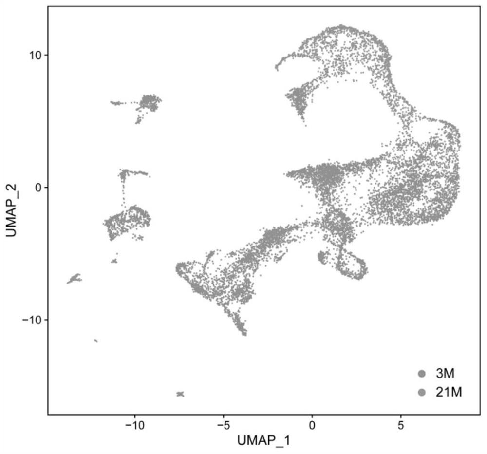
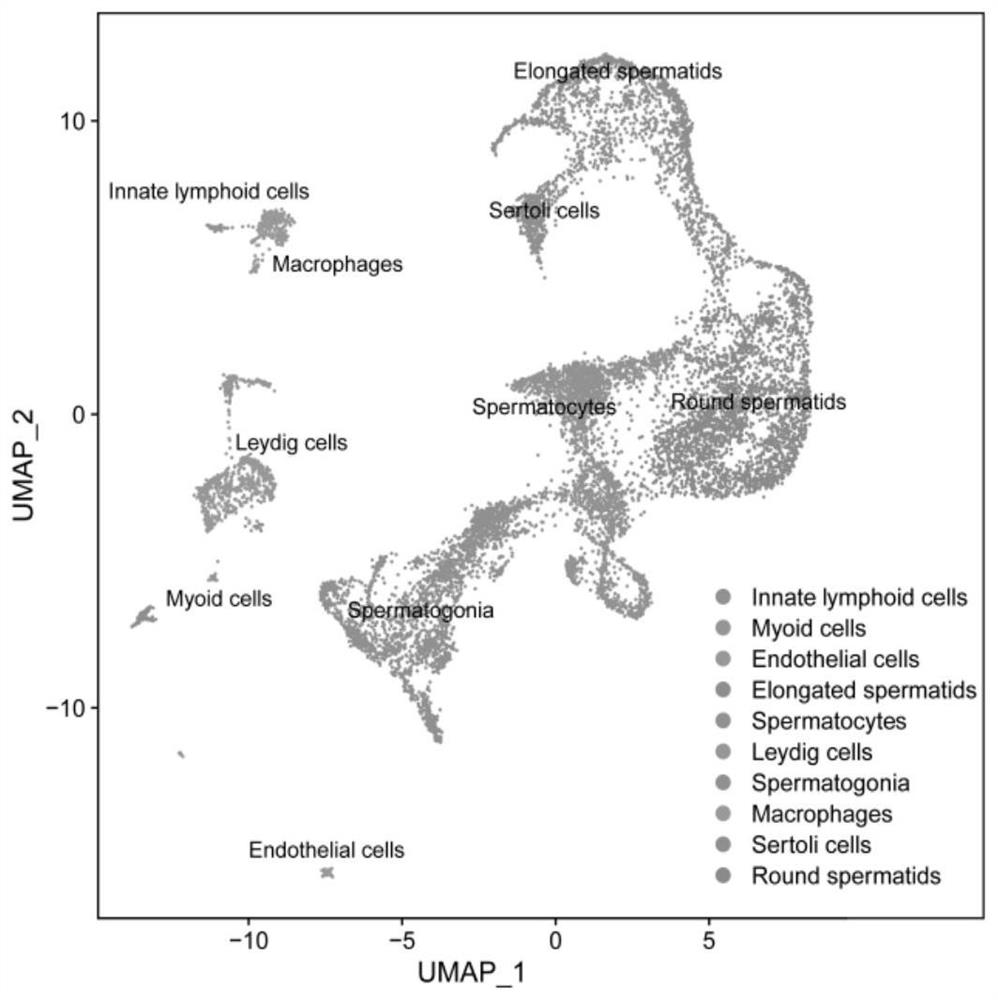
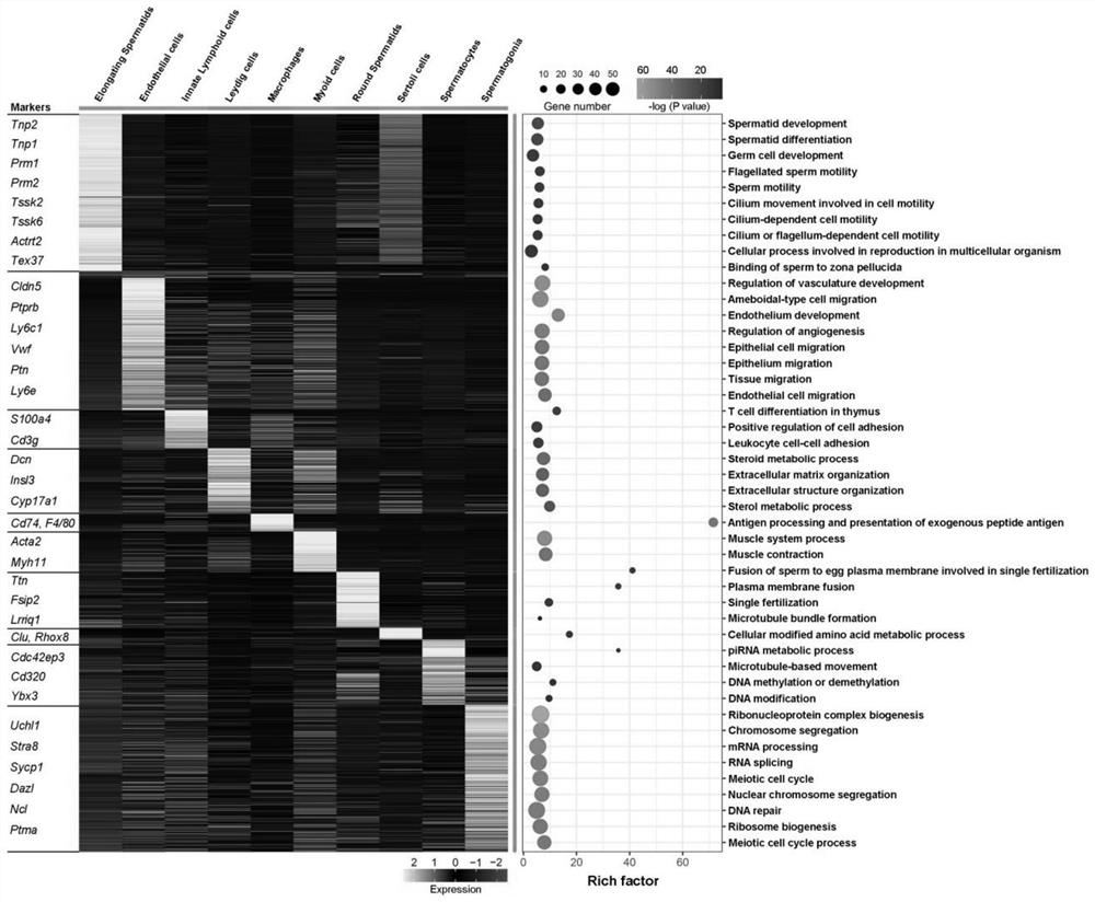
[0042] In this embodiment, the 3-month-old (3M) and 21-month-old (21M) mouse testicular tissue cells extracted in Example 1 were subjected to cell sequencing, and the distribution of cells in the testis of mice of different ages, the overall cell grouping and the number of cells in the testis were observed. Expression of macrophage markers in testicular cells. Experimental results such as Figure 1 ~ Figure 4 shown. Depend on figure 1 It can be seen that 7524 testicular cells were captured in the 3M group, and 3597 testicular cells were captured in the 21M group. There was no significant change in the ratio of cell numbers between the two. Depend on figure 2It can be seen that after removing the haploid, the main cell groups in the testes of the 3M group and the 21M group include innate lymphocytes, peritubercular myoid cells, endothelial cells, Leydig cells, macrophages, spermatogonia, and spermatocytes , elongated sperm, round sperm and Sertoli cells. In addition, the...

Embodiment 3

[0044] In this example, the 3-month-old (3M) and 21-month-old (21M) mouse testicular macrophages extracted in Example 1 were sequenced to observe the expression of CD74 and various protein markers in mouse macrophages of different ages . Experimental results such as Figure 5 ~ Figure 7 shown. Depend on Figure 5 ~ Figure 7 It can be seen that the expression level of CD74 in the testicular macrophages of 3-month-old (3M) and 21-month-old (21M) mice is higher than that of other proteins.


PUM


Abstract

The invention discloses a sorting method of macrophages in testis, and relates to the technical field of biological cells. The invention provides an application of CD74 as a protein marker in macrophage sorting. Through a large number of experimental screening, the inventor finally finds that compared with existing common macrophage protein markers including F4 / 80, CD206, CD86, CD68, CD14, CD163 and CD11b, the testis macrophage protein marker CD74 disclosed by the invention has wider expression in a testis macrophage group, and shows the strongest testis specificity; by adopting the sorting method for the macrophages in the testis, the testis macrophages can be accurately and efficiently separated.

Description

technical field [0001] The invention relates to the technical field of biological cells, in particular to a method for sorting intratesticular macrophages. Background technique [0002] Macrophages are immune cells that are part of the mononuclear phagocyte system, whose main role is to engulf dead cells, cell debris, pathogens, and other foreign matter. In addition, they have signaling and regulatory functions. Macrophages have strong tissue specificity and plasticity, and can change their phenotype in response to environmental signals. Currently, macrophages have been extensively and intensively studied in the fields of aging, immunity, development, tissue repair, and cancer. Macrophages exist in many tissues of the human body, and it is not a group of cells with uniform function, but a group of cells with rich heterogeneity. Different macrophage subpopulations have distinct gene expression profiles and cellular functions. In order to better understand these difference...

Claims


Application Information

Patent Timeline
IPC IPC(8): C12N5/0786G01N33/569
CPCC12N5/0645G01N33/56977C12N2509/10C12N2509/00G01N2333/70539G01N2469/10
Inventor 谢云邓春华王竹孙祥宙姚嘉慧涂响安
Owner THE FIRST AFFILIATED HOSPITAL OF SUN YAT SEN UNIV
Learn More