
Cerebral hemorrhage CT image classification method based on image sequence analysis

An image sequence and CT image technology, applied in the field of medical image analysis, can solve the problems of ignoring the context, the complex acquisition of fine marks for recognition and segmentation tasks, and the lack of special attention of important features, so as to achieve the effect of improving the diagnostic ability

Pending Publication Date: 2022-05-31
NANJING UNIV OF AERONAUTICS & ASTRONAUTICS
View PDF0 Cites 2 Cited by
  • Summary
  • Abstract
  • Description
  • Claims
  • Application Information

AI Technical Summary

Problems solved by technology

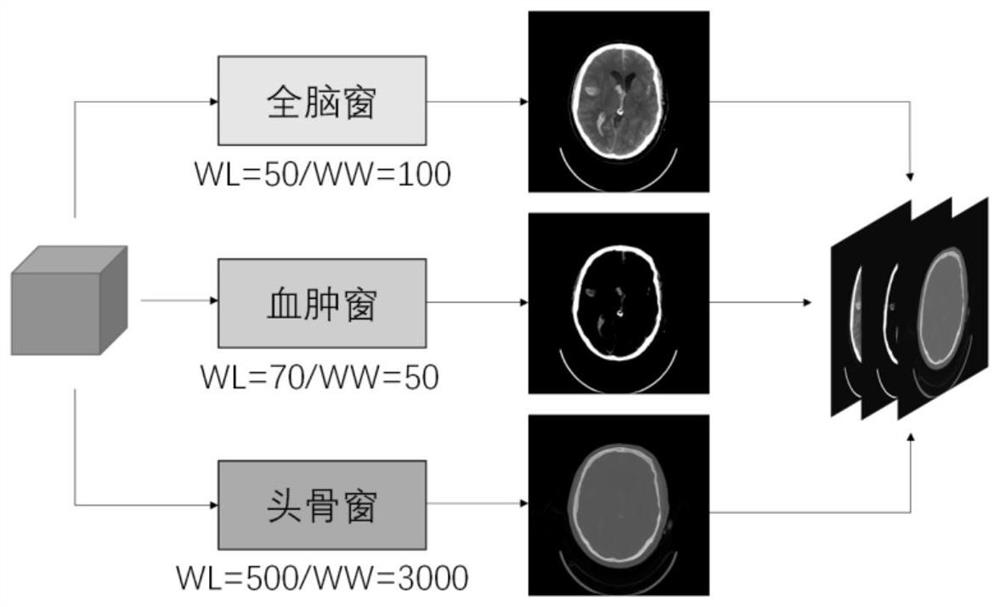
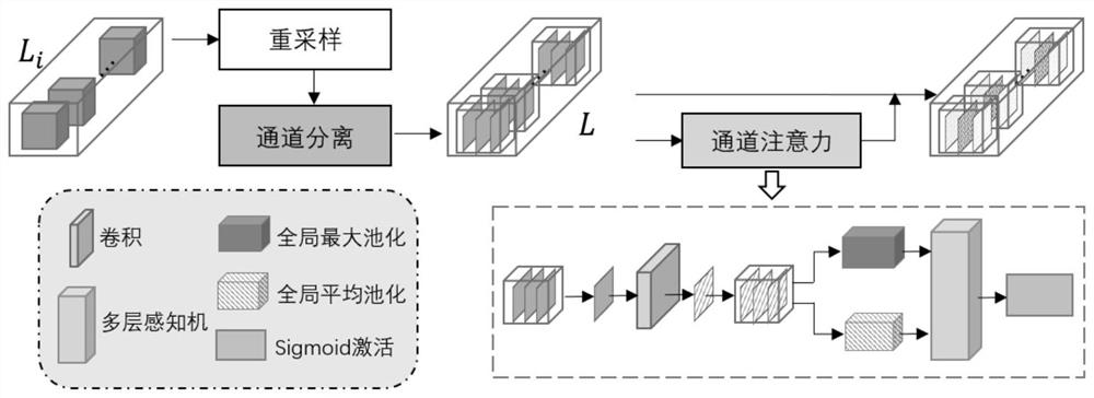
[0004] Purpose of the invention: The purpose of the invention is to solve some problems in the existing work related to the diagnosis of cerebral hemorrhage based on brain CT images, including: the acquisition of refined markers in recognition and segmentation tasks is complicated, and the diagnostic method for a single scan layer ignores The contextual relationship contained in the entire sample scanning sequence, the setting of the window level and window width is easy to lose information during the graphical display of DICOM data, and the model method lacks special attention to important features, etc.; provides a method based on image sequence analysis The CT image classification method of cerebral hemorrhage has its significance through artificial intelligence-assisted clinical diagnosis of cerebral hemorrhage; specifically, the present invention solves the above problems to a certain extent; wherein, the image is improved by multi-window and focusing on the overall scanning layer sequence. The degree of utilization of information, the introduction of channel attention and spatial attention mechanism convolutional long short term memory (Convolutional long short term memory, CLSTM) network to extract and differentiate the features in the multi-channel image sequence, based on the extraction of scan samples Reasonable global feature representation, training an end-to-end cerebral hemorrhage classification model, intelligently diagnosing the occurrence of cerebral hemorrhage and distinguishing the subtypes of cerebral hemorrhage, thus providing assistance for clinical screening of cerebral hemorrhage diseases

Method used


Image

  • Cerebral hemorrhage CT image classification method based on image sequence analysis
  • Cerebral hemorrhage CT image classification method based on image sequence analysis
  • Cerebral hemorrhage CT image classification method based on image sequence analysis

Examples


Embodiment Construction

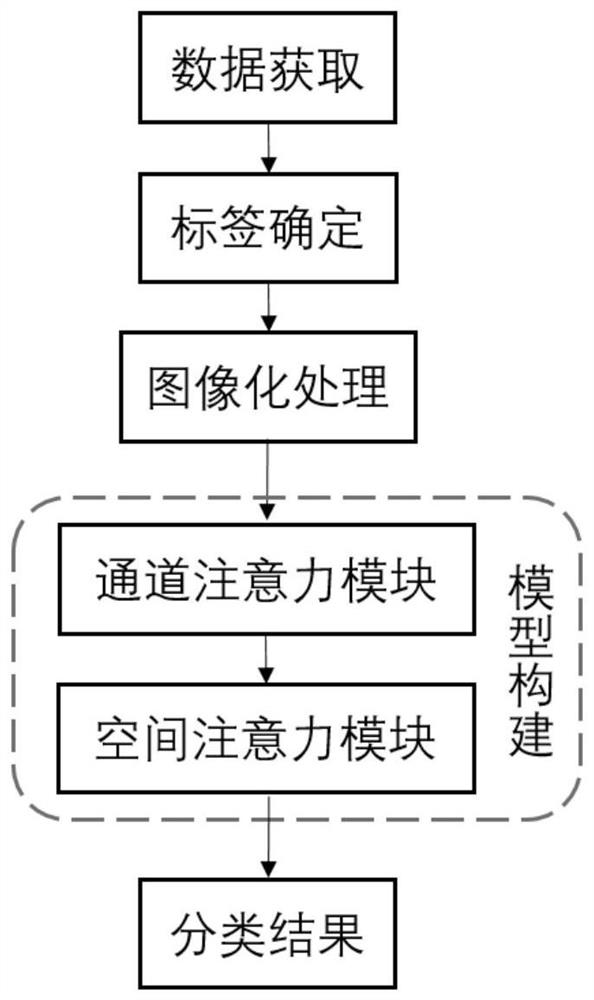
[0028] The present invention will be further described below in conjunction with the accompanying drawings and embodiments.

[0029] As shown in the figure, a method for classifying CT images of cerebral hemorrhage based on image sequence analysis according to the present invention first classifies CT images of cerebral hemorrhage; specifically:

[0030]Determine whether cerebral hemorrhage occurs from the brain CT scan, that is, judge whether the brain CT scan result is positive for cerebral hemorrhage or negative for cerebral hemorrhage; for positive samples of cerebral hemorrhage, identify the subtype of cerebral hemorrhage, that is, judge whether the brain CT scan result is positive or negative. Whether the cerebral hemorrhage shown is intraventricular hemorrhage, parenchymal hemorrhage, subarachnoid hemorrhage, epidural hemorrhage, or subdural hemorrhage.

[0031] Furthermore, a method for classifying CT images of cerebral hemorrhage based on image sequence analysis, the ...


PUM


Abstract

The invention discloses a cerebral hemorrhage CT image classification method based on image sequence analysis. The method belongs to the technical field of medical image analysis, and the operation process comprises the steps of brain CT image data acquisition, data imaging processing, model construction and classification method implementation. The method comprises the following specific operation steps: acquiring a brain CT scanning result to obtain brain CT image original data; performing imaging processing on the original data of the brain CT image to form a three-channel image sequence as the input of a subsequent model; constructing a model comprising a channel attention module and a space attention module to extract features of the three-channel image sequence; according to the method, the model is trained to optimize parameters, cerebral hemorrhage positive is recognized, cerebral hemorrhage subtypes are distinguished, cerebral hemorrhage CT images are classified, CT scanning image information can be fully utilized by the model, attention to key information in an image sequence is improved, the judgment process of the whole method conforms to the diagnosis process of doctors in a real scene, and the diagnosis efficiency is improved. And the cerebral hemorrhage classification effect can be improved.

Description

technical field [0001] The invention belongs to the technical field of medical image analysis, and relates to a method for classifying CT images of cerebral hemorrhage based on image sequence analysis. Background technique [0002] With the continuous development of computer-related technologies, artificial intelligence methods are increasingly used in various fields, and medical image analysis related work based on machine learning and deep learning methods can improve the diagnostic efficiency of clinically related diseases and reduce the workload of doctors. ;In the clinical diagnosis of cerebral hemorrhage, it is often based on the patient's brain computerized tomography (CT) image results. This medical image performs a predetermined interval of tomographic scans on the brain and obtains multiple image layers. The scan time is fast and the image With sufficient clarity, non-contrast-enhanced plain scans are generally preferred in clinical CT examinations; cerebral hemorr...

Claims


Application Information

Patent Timeline
IPC IPC(8): G06T7/00G06K9/62G06V10/764G06V10/774G06V10/82G06N3/04G06N3/08
CPCG06T7/0012G06N3/08G06T2207/10081G06T2207/20081G06T2207/20084G06T2207/30016G06N3/044G06F18/24G06F18/214
Inventor 张道强祝小惟
Owner NANJING UNIV OF AERONAUTICS & ASTRONAUTICS
Learn More