Plasticized specimen for displaying anatomical characteristics of fascia fiber construction and anatomy method

A technology of anatomy and fascia, applied in teaching models, preservation of human or animal bodies, instruments, etc., to achieve high social and economic benefits and wide application value

Pending Publication Date: 2022-06-03
DALIAN MEDICAL UNIVERSITY
View PDF2 Cites 0 Cited by
  • Summary
  • Abstract
  • Description
  • Claims
  • Application Information

AI Technical Summary

Problems solved by technology

At present, there are no specimens and techniques that can comprehensively display the anatomical chara...

Method used


Image

  • Plasticized specimen for displaying anatomical characteristics of fascia fiber construction and anatomy method
  • Plasticized specimen for displaying anatomical characteristics of fascia fiber construction and anatomy method
  • Plasticized specimen for displaying anatomical characteristics of fascia fiber construction and anatomy method

Examples


Embodiment 1

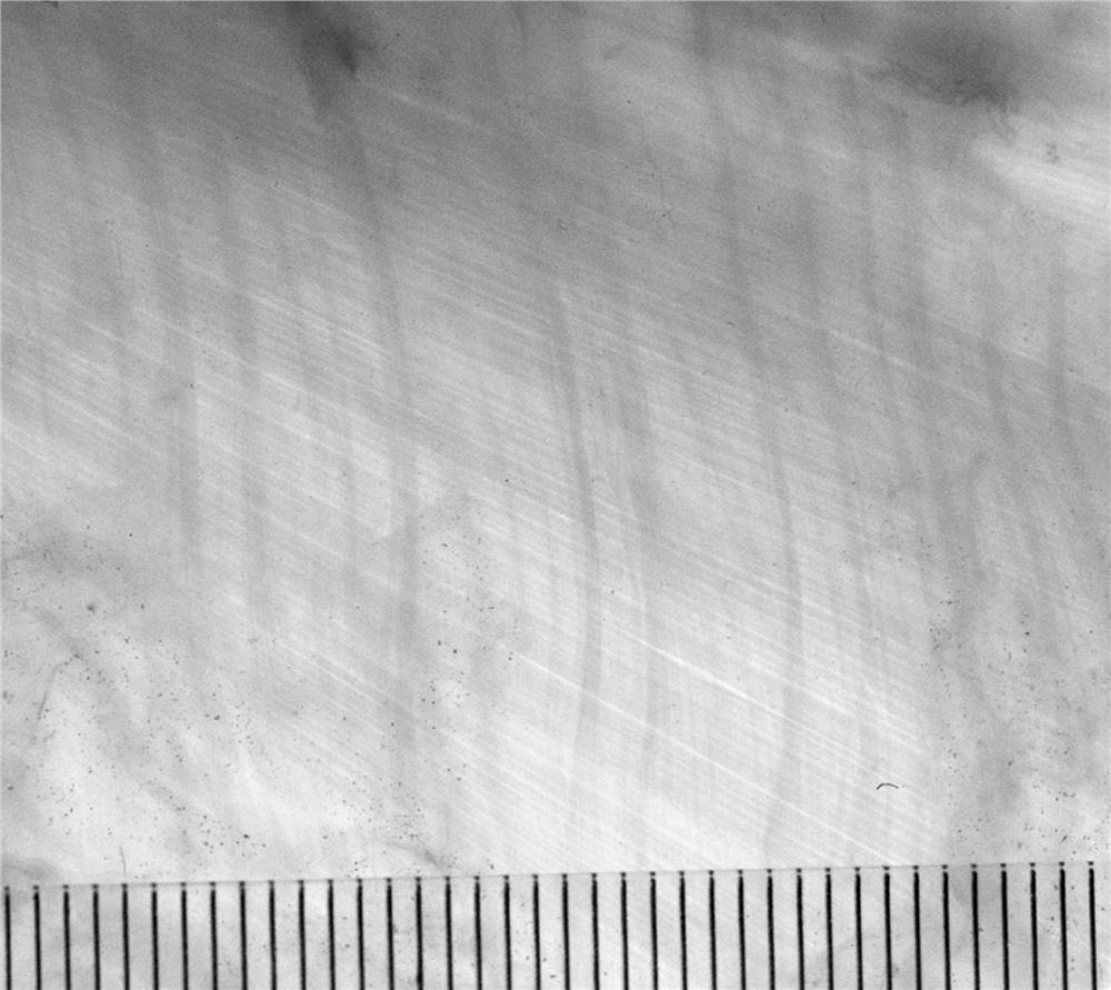
An anatomical method for displaying the anatomical features of fascial fibers in this embodiment includes the following steps:

1. Anatomy

After the iliotibial band around the knee joint that has been separated from the human body is fixed with formalin solution, the fascia and its related structures in the target area are dissected to obtain fascia specimens.

[0022] bleach

The fascia specimens were bleached with 5% hydrogen peroxide and observed once an hour to prevent over-bleaching. After about 24 hours, they were soaked and rinsed with small running water for 1 day.

[0023] dehydration

Expand the specimen and cut through the non-primary observation area to flatten the fascia specimen. Specimens were fixed using splint grids and dehydrated by immersion in acetone liquid at -25°C and -15°C, respectively. First dehydration, 90% acetone at -25°C, 3 days. Second dehydration, 95% acetone at -15°C, 4 days.

[0024] degreased

3 times degreasing in acetone liquid at ro...

Embodiment 2

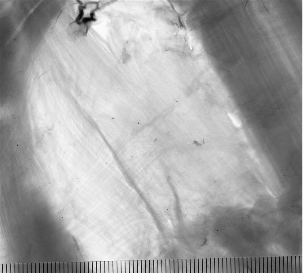
Referring to Embodiment 1, the difference from Embodiment 1 is that the parts that have been separated from the human body or large animals in this embodiment are the ligaments and fascia around the knee joint.

[0029] The produced plastinated specimens are shown in figure 2 shown.

Embodiment 3

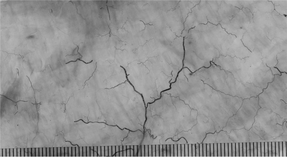
Referring to Embodiment 1, the difference from Embodiment 1 is that the part that has been separated from the human body or large animal in this embodiment is the middle layer of the thoracolumbar fascia.

[0031] The produced plastinated specimens are shown in image 3 shown.


PUM


Abstract

The invention discloses a plasticized specimen for displaying anatomical characteristics of fascia fiber construction and a dissecting method, and the method comprises the steps: S1, after a part separated from a human body or a large animal is subjected to anticorrosion fixation, dissecting and sampling of a fascia in a target area is carried out, and a fascia specimen is obtained; s2, bleaching the fascia specimen with hydrogen peroxide, and then soaking and washing the fascia specimen with small flowing water; s3, unfolding the specimen, and flattening the fascia specimen; fixing the specimen by using a splint grid, and respectively immersing the specimen into two acetone liquids at different temperatures for dehydration; s4, degreasing in normal-temperature acetone liquid with different concentrations for multiple times; s5, preparing a glass clamping piece, and adding the glass clamping piece into a P45 mixed solution for full embedding; completely infiltrating the P45 mixed solution in a low-temperature negative-pressure state, and hardening the P45 mixed solution in a water bath box; and S6, taking out the transparent and hardened fascia specimen, and cutting, trimming and forming the fascia specimen. The anatomical characteristics of fiber construction of fascia of a certain part of a human body or a large animal are comprehensively displayed on the macroscopic level.

Description

technical field [0001] The invention relates to the field of biotechnology, in particular to a plasticized specimen and an anatomical method for displaying the anatomical features of fascia fibers. Background technique [0002] Existing anatomical specimens and anatomical methods have limitations in the display of fascial fibrous structures. The most common formalin-fixed gross anatomical specimens are able to observe the overall morphology of the fascia, but not the fibrous structure within the fascia. Microscopic histological sectioning and staining methods, through serial sectioning, can display a small range of fascial fiber structures, but cannot display the anatomical features of a large range of fascial fiber structures. In addition, the formalin-fixed tomographic specimen can only display the position information of the fascia on the specimen section. The newly emerged tomographic plastination specimens can only show the anatomical features of the fascial fibers on...

Claims


Application Information

Patent Timeline
IPC IPC(8): A01N1/00G09B23/30
CPCA01N1/00G09B23/306G09B23/30
Inventor 于胜波张健飞孟宪荣迟彦艳隋鸿锦韩建马学伟朱高麟
Owner DALIAN MEDICAL UNIVERSITY
Why Eureka
Try Eureka