Retina image blood vessel segmentation method based on improved U-Net network

A retina and network technology, applied in the field of image processing, can solve the problem of inaccurate segmentation of segmentation technology

Pending Publication Date: 2022-08-09
GUILIN UNIVERSITY OF TECHNOLOGY
View PDF0 Cites 1 Cited by
  • Summary
  • Abstract
  • Description
  • Claims
  • Application Information

AI Technical Summary

Problems solved by technology

[0005] In order to solve the problem of imprecise segmentation of the existing segmentation technology, the p

Method used


Image

  • Retina image blood vessel segmentation method based on improved U-Net network
  • Retina image blood vessel segmentation method based on improved U-Net network
  • Retina image blood vessel segmentation method based on improved U-Net network

Examples


Embodiment Construction

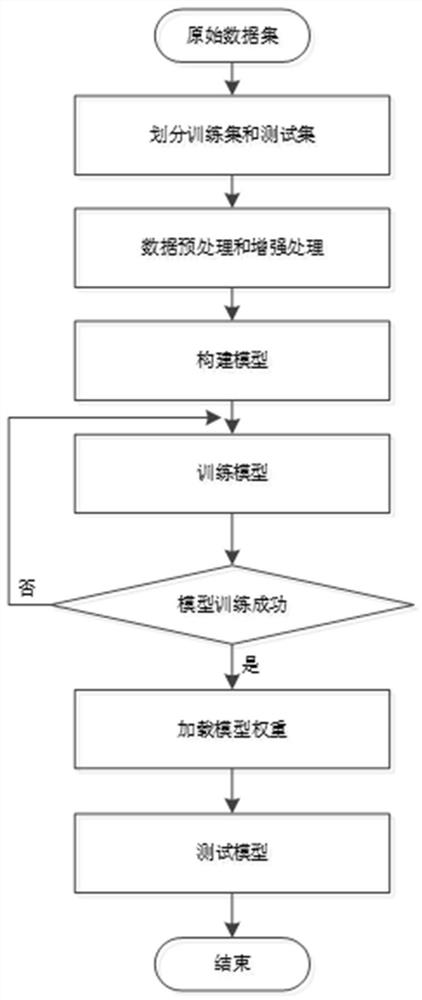
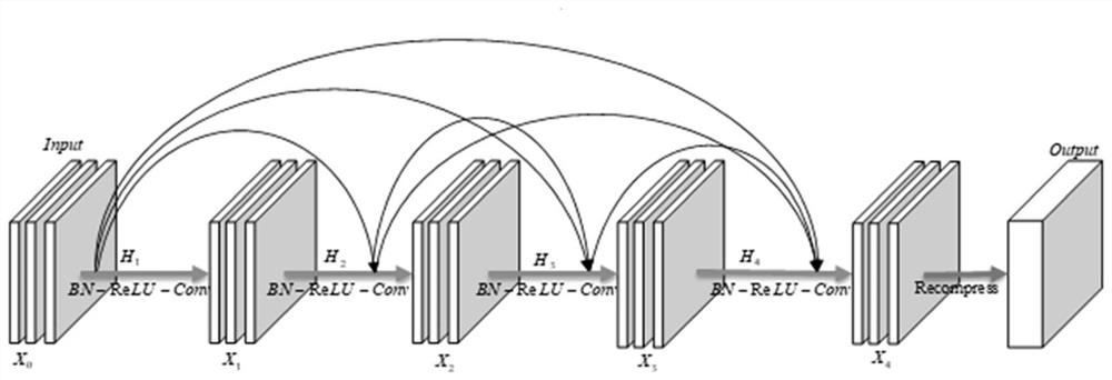
[0026] According to an embodiment of the present invention, a retinal image blood vessel segmentation method based on an improved U-Net network is proposed. The U-Net framework is simplified, a symmetrical 3-pass encoding-decoding structure is adopted, traditional convolution is optimized, an attention mechanism is introduced, and finally the accurate segmentation effect of the model is achieved. The present invention will be further described in detail below with reference to the drawings and specific examples. The blood vessel segmentation flowchart of the present invention is as follows: figure 1 shown. The retinal image blood vessel segmentation method based on the improved U-Net network of the present invention specifically comprises the following steps:

[0027] Step 1: Obtain the public color retinal fundus blood vessel segmentation dataset DRIVE;

[0028] Step 2: Randomly divide the original data set, take 20 for the validation set and 20 for the test set;

[0029] ...


PUM


Abstract

The invention provides a retinal vessel segmentation method based on an improved U-Net network. Image enhancement is performed on a color eye fundus image, so that the contrast ratio between a blood vessel and a background in the image is improved, and a training data set is amplified. A U-Net encoder-decoder structure is used as a basic segmentation framework, a dense convolution block and a CDBR layer structure are designed to replace a traditional convolution block, learning of multi-scale feature information is achieved, and the feature extraction capacity of the model is improved. Meanwhile, an attention mechanism is introduced at a jump connection part of the model, so that the model is enabled to allocate weights again, the importance degree of a feature channel is adjusted, noise is suppressed, the problem of blood vessel information loss in an up-sampling process at a decoder end is solved, and a GAB-D2BUNet network model is constructed based on the above technologies. According to the method, an internationally disclosed retina fundus blood vessel data set DRIVE is adopted for training, and finally the optimal segmentation model is reserved to verify the segmentation performance of the model. The retina fundus blood vessel segmentation method achieves the task of accurately segmenting the retina fundus blood vessel, and has better segmentation performance.

Description

technical field [0001] The invention relates to an image segmentation method using deep learning, in particular to a retinal image blood vessel segmentation method based on an improved U-Net network, and belongs to the field of image processing. Background technique [0002] Diabetic retinopathy is a complication of diabetes with high morbidity and blindness rates. Diabetic retinopathy is a retinal disease caused by elevated blood sugar. High blood sugar levels in the body can cause blockage or damage to the tiny retinal blood vessels that nourish the retina. In order to maintain eye nutrition metabolism, the retina will breed new tiny blood vessels. Fragile and prone to oozing and bleeding of vascular material. Over time, patients will experience blurred vision and, in more serious cases, blindness. According to the World Health Organization, about 220 million people worldwide are affected by diabetes. Because the morphological changes of retinal blood vessels directly r...

Claims


Application Information

Patent Timeline
IPC IPC(8): G06T7/00G06T7/11G06T5/00G06N3/04G06N3/08G06T5/40
CPCG06T7/0012G06T7/11G06T5/002G06N3/08G06T5/40G06T2207/20021G06T2207/20081G06T2207/20084G06T2207/30041G06T2207/30101G06N3/045Y02T10/40
Inventor 程小辉李贺军黎辛晓
Owner GUILIN UNIVERSITY OF TECHNOLOGY
Why Eureka
Try Eureka