Analytical test device and method
A technology for analyzing assay devices and analytes, applied in measuring devices, analytical materials, biological material analysis, etc., can solve the problems of large amount of labeling, large detection area, increased assay time, etc., and achieve the effect of rapid and effective flow
- Summary
- Abstract
- Description
- Claims
- Application Information
AI Technical Summary
Problems solved by technology
Method used
Image
Examples
example 1
[0303] Whole blood CK-MB test
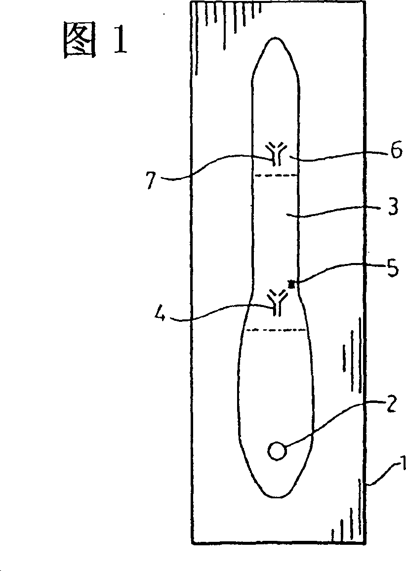
[0304]1A) On a polyester supported nitrocellulose film (Gerbermenbrane GmbH, Gerbershausen, Germany), the contour shown in Figure 4 was drawn with Paint Marker 751 Yellow (Edding AG, Ahrensburg, Germany). Capture test lines were prepared with 13 mg / ml streptavidin in water (Streptavidin, poly, from Micrsoft GmbH, Benried, Germany). Controls were prepared with 80 μl of a 4% (wt / vol) sucrose solution (from Sigma-Aldrich GmbH, Steinheim, Germany), 10 μl of water, and 10 μl of a 1 mg / ml recombinant CK-MB solution (from Spectral Diagnostics, Toronto, Canada). Test line. After drying, octyl-beta-D-gluco-pyranoside (Octyl-beta-D-Gluco-Pyranoside) (from Sigma-Aldrich GmbH in Steinheim, Germany), 1 : 30 dilutions of Kasein-Bindemittel (from H. Schmineke & Co., Erkrath, Germany) and 30 mM 1,4-piperazinediethanesulfonic acid (from Sigma-Aldrich GmbH, Steinheim, Germany) with a final pH value of 6.2 The blocking solution impregnates the ...
example 2
[0318] Comparison of semicircular and rectangular CK-MB assays
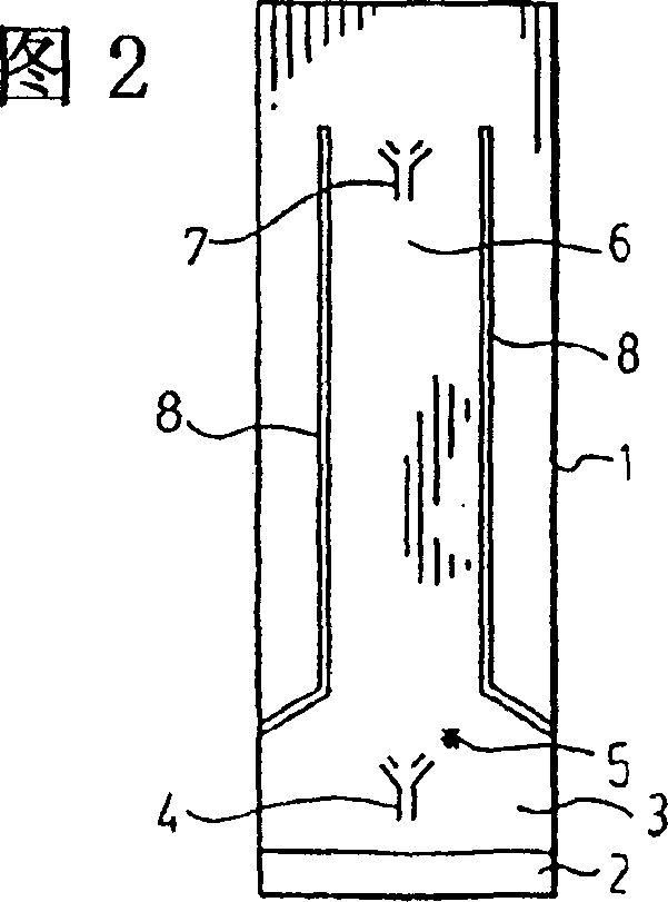
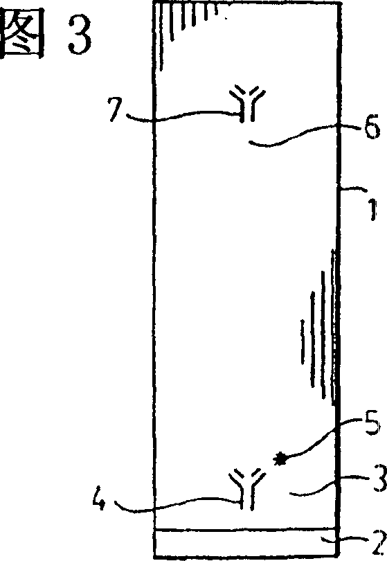
[0319] To illustrate the concept of generality, the specimen was brought into a semicircular area (circular section) (Fig. 4 and Figure 5 ) and the specimen enters the rectangular area (ie Figure 9 Compare with side 3) of Figure 10. The assay area (contour area) was the same in both cases. All the steps are the same as in Example 1) except for the outline shape and the direction of blood entry.
[0320] rCKMB Circular part Rectangular
[0321] ng / ml Signal Assay time Signal Assay time
[0322] 0 - 6.5 minutes - 7.5 minutes
[0323] 20 ++ 7.0 minutes ++ 7.5 minutes
example 3
[0325] Semi-circular area - 3 analytes - one detection area
[0326] Prepare the assay as in Example 1B), but in addition to having the CKMB antibody capture line, the TNI antibody capture line and the myoglobin antibody capture line.
[0327] TNI capture: 13mg / ml polyclonal goat TNI
[0328] CKMB Capture: lrCKMB-28 at 13mg / ml
[0329] Myoglobin capture: 13mg / ml polyclonal rabbit myoglobin
[0330] All antibodies were from Spectral Diagnostics, Toronto.
[0331] The conjugated alloys corresponding to the 3 analytes were from British BiocellIntern.. from Cardiff, UK:
[0332] TNI-gold-a: 40nm gold sol filled with 8 μg / ml of 81-7 antibody (OD10)
[0333] TNI-gold-b: 40nm gold sol filled with 16μg / ml 21-14 antibody (OD10)
[0334] Myoglobin-gold: 15nm gold sol filled with 90μg / ml 2Mb-295 antibody (OD10)
[0335] CK-MB gold: 40nm gold sol filled with 22μg / ml 5CKMB-6 (OD10)
[0336] All antibodies were from Spectral Diagnostics, Toronto.
[0337] The TNI-gold b...
PUM
| Property | Measurement | Unit |
|---|---|---|
| particle diameter | aaaaa | aaaaa |
| aperture size | aaaaa | aaaaa |
| composition ratio | aaaaa | aaaaa |
Abstract
Description
Claims
Application Information
 - R&D
- Intellectual Property
- Life Sciences
- Materials
- Tech Scout
- Unparalleled Data Quality
- Higher Quality Content
- 60% Fewer Hallucinations
Browse by: Latest US Patents, China's latest patents, Technical Efficacy Thesaurus, Application Domain, Technology Topic, Popular Technical Reports.
© 2025 PatSnap. All rights reserved.Legal|Privacy policy|Modern Slavery Act Transparency Statement|Sitemap|About US| Contact US: help@patsnap.com


