Method for digita image reducing angiography using primary stereo data

A technology of angiography and digital subtraction, applied in image data processing, equipment for radiological diagnosis, application, etc.

Active Publication Date: 2004-10-20
SIEMENS HEALTHCARE GMBH
View PDF0 Cites 13 Cited by
  • Summary
  • Abstract
  • Description
  • Claims
  • Application Information

AI Technical Summary

Problems solved by technology

[0003] However, this known technique has the disadvantage that until now two radiographs had to be produced in order to carry out the subtraction angiography, a filling photograph and a mask

Method used


Image

  • Method for digita image reducing angiography using primary stereo data

Examples


Embodiment Construction

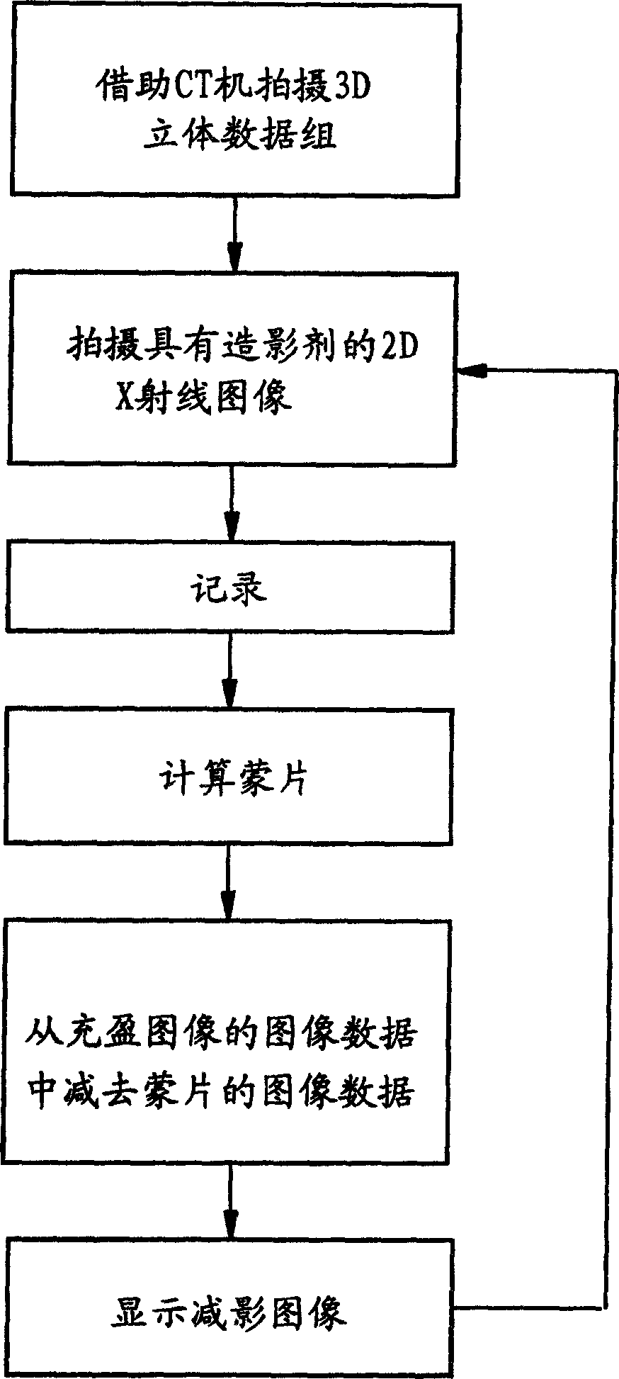
[0013] figure 1 An example flowchart of a method of the present invention for displaying blood vessels of a patient is shown. First, a 3D volumetric data record of the patient or at least one body region of the patient is generated. The data set can be acquired with an x-ray computed tomography system or an angiography system. It is important here that no contrast agent is injected into the blood vessel during the 3D recording, so that the anatomical background in any direction of projection can be reproduced from the volume data. The 3D volumetric data is stored by known means and used for later further processing.

[0014] When carrying out the subtraction angiography according to the method according to the invention, the physician takes a 2D x-ray of the body region of interest in the desired viewing or projection direction, in which the vessels are filled with contrast agent, in order to obtain a filling image.

[0015] The 3D stereoscopic data set and the 2D X-ray pho...


PUM


Abstract

In a method to implement digital subtraction angiography, an image of structures (in particular vessels) of a body region is generated by subtraction of image data of a first 2D x-ray image, that is created without enrichment of the structures with contrast agent, from image data of a second 2D x-ray image of the body region that is acquired with contrast agent enrichment of the structures. The image data of the first 2D x-ray image are calculated from a 3D volume dataset of the body region. The x-ray dose applied in the subtraction angiography thus can be reduced.

Description

technical field [0001] The invention relates to a method for carrying out digital subtraction angiography (Subtraktions angiographie), wherein by subtracting from the image data of the second 2D X-ray images of the structure of the body region, especially the blood vessels, taken when filled with contrast agent The image data of the first 2D x-ray image produced when the structure of the body region was not filled with contrast agent is used to generate an image of the structure of the body region. Background technique [0002] Subtraction angiography is used in particular to visualize the blood vessels of the body in order to be able to identify possible vascular abnormalities such as narrowing (stenosis) or widening of blood vessels (aneurysm) in the visualization. To improve the visibility of blood vessels in an X-ray image, a contrast agent is added to the blood before the X-ray image is taken, so that the blood vessels stand out clearly from the background in the X-ray ...

Claims


Application Information

Patent Timeline
Patent Type & Authority Applications(China)
IPC IPC(8): A61B6/00A61B6/03G06T5/50G06T7/00
CPCG06T2207/10081A61B6/481G06T2207/10124G06T7/0038A61B6/032G06T5/50G06T2207/30101A61B6/504G06T7/38
Inventor 乔基姆·霍恩格尔
Owner SIEMENS HEALTHCARE GMBH
Why Eureka
Try Eureka