Real time method for detecting nucleic acid ligase reaction and nucleic acid ligase chain reaction

A ligase reaction, real-time detection technology, applied in the field of molecular biology

Inactive Publication Date: 2005-06-15
HUNAN UNIV
View PDF0 Cites 5 Cited by
  • Summary
  • Abstract
  • Description
  • Claims
  • Application Information

AI Technical Summary

Problems solved by technology

[0005] The invention aims to develop a real-time fluorescent nucleic acid ligase reaction and nucleic acid ligase chain reaction detection method, which is applied to the analysis and research of nucleic acid single nucleotide polymorphism, solves the shortcomings of the existing gene analysis technology, and promotes related research development

Method used


Image

  • Real time method for detecting nucleic acid ligase reaction and nucleic acid ligase chain reaction
  • Real time method for detecting nucleic acid ligase reaction and nucleic acid ligase chain reaction
  • Real time method for detecting nucleic acid ligase reaction and nucleic acid ligase chain reaction

Examples


Embodiment 1

[0019] Embodiment 1 (verification experiment of detection method principle)

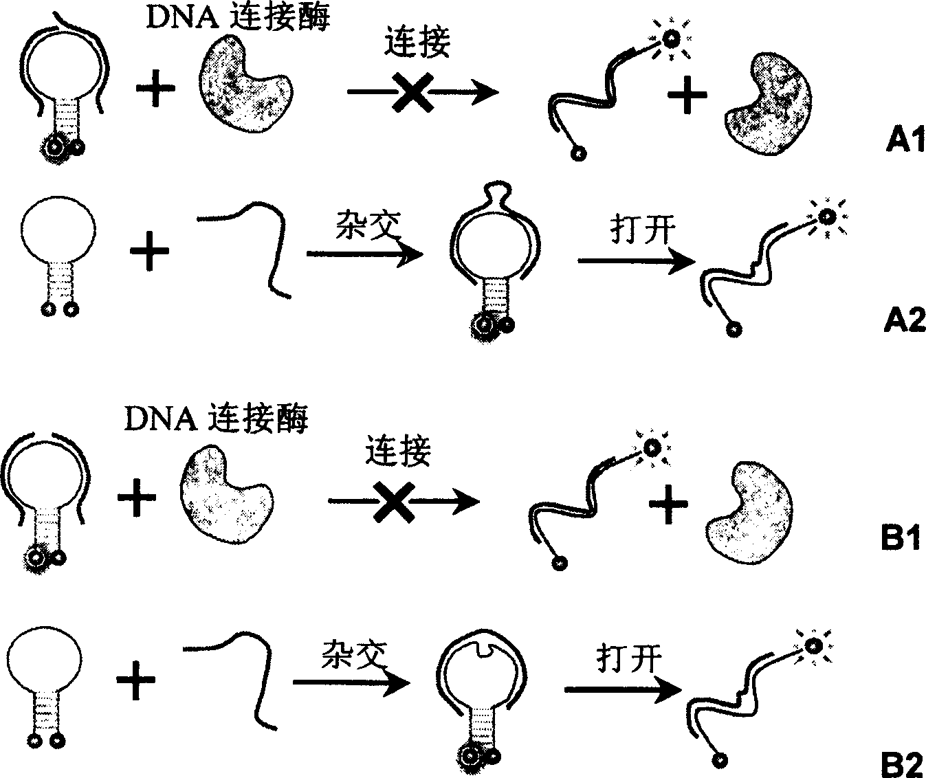
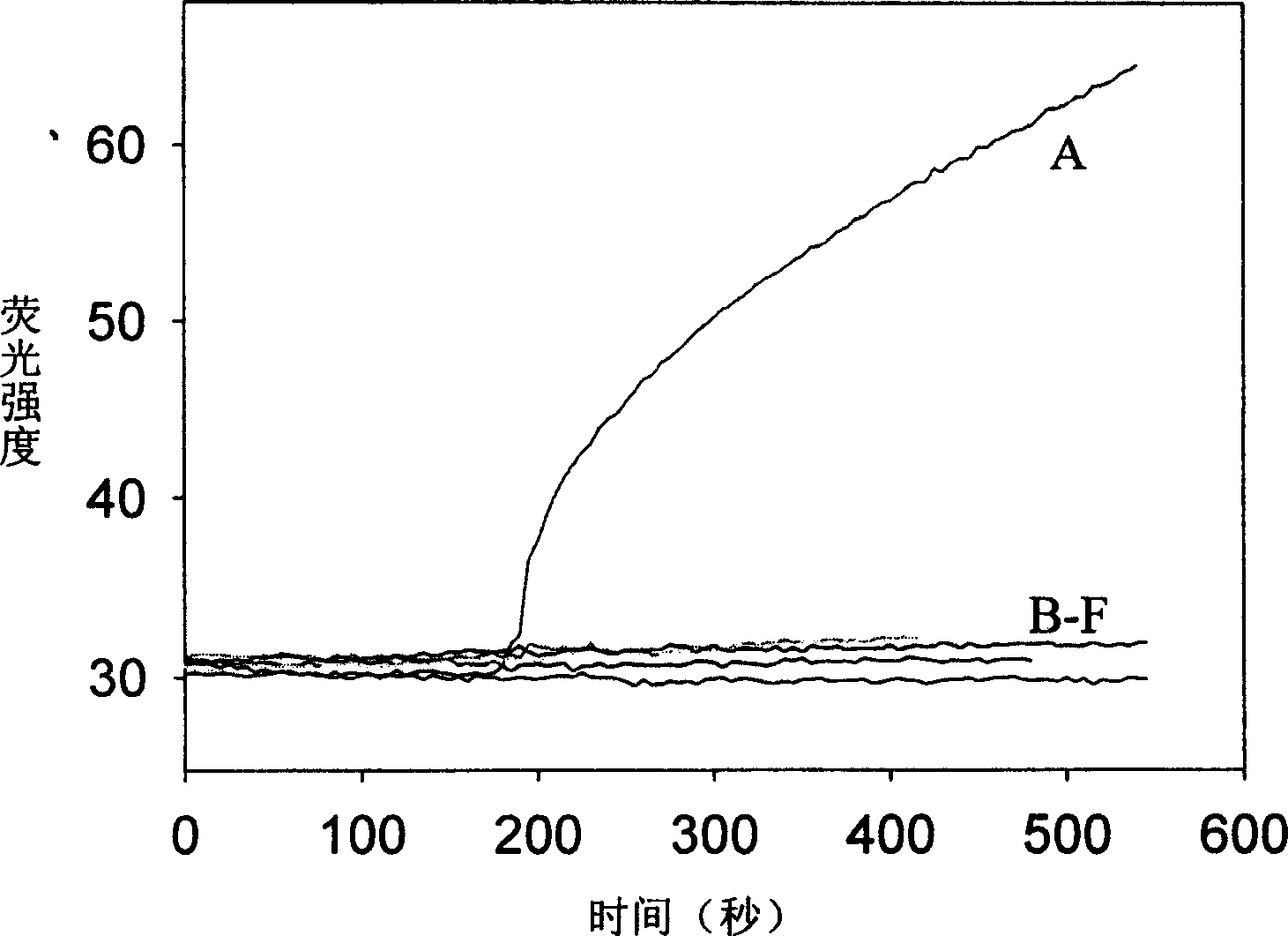
[0020] Six samples were prepared in the experiment, in 200uL sample buffer (including 20mM Tris-HCl (pH=7.6), 25mM KAc, 10mM Mg(Ac) 2 , 10mM DTT, 1mM NAD, 0.1% Triton-X100) and MB with a final concentration of 200nM, in addition, different DNA primers were added to each sample: (A) Primer1+Primer4; (B) Primer1+Primer5; (C) Primer2 +Primer5; (D) Primer1+Primer6; (E) Primer3+Primer4; (F) Primer3+Primer6; where the final concentration of Primer1 to Primer6 is 200 nM. The sample was placed in a F2500 fluorometer, and the sample temperature was kept constant at 45 degrees Celsius. Then the fluorescence intensity of the sample was detected with the parameters of excitation wavelength 497nm and emission wavelength 521nm. After the fluorescence intensity is stable, add 10U of Taq ligase, monitor and record the fluorescence intensity of the sample in real time, the results are as follows: figure 2 shown. ...

Embodiment 2

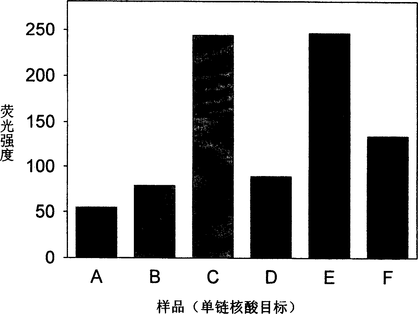
[0027] Embodiment 2 (real-time LDR method is to the analysis of single-stranded nucleic acid object)

[0028] Six samples were prepared in the experiment, in 200uL sample buffer (including 20mM Tris-HCl (pH=7.6), 25mM KAc, 10mM Mg(Ac) 2 , 10mM DTT, 1mM NAD and 0.1% Triton-X100) were added with 30U of Taq ligase and MB with a final concentration of 200nM, except for one MB control sample A without DNA samples, other samples contained (B) PrimerA +PrimerB; (C) MutantDNA1+PrimerA+PrimerB; (D) NormalDNA1+PrimerA+PrimerB; (E) MutantDNA1+PrimerC+PrimerD; (F) NormalDNA1+PrimerC+PrimerD; is 200nM, and the concentration of template DNA is 10nM. Put these 5 samples in a PCR machine, set the program for LDR temperature cycle, stay at 45 degrees Celsius for 5 minutes for ligation amplification, raise the temperature to 80 degrees Celsius for 3 minutes to melt the DNA, and do 25 cycles in total. Then, the fluorescence intensity of the sample was detected with a F2500 fluorescence instrum...

Embodiment 3

[0030] Embodiment 3 (real-time LDR method is to the analysis of double-stranded nucleic acid object)

[0031]In the experiment, 6 samples were prepared, and 30U of Taq ligase and MB with a final concentration of 200nM were added to 200uL sample buffer (same as Example 2). Except for the control sample A of one MB without adding other DNA samples, other samples (B) PrimerA+PrimerB; (C) MutantDNA1+MutantDNA2+PrimerA+PrimerB; (D) NormalDNA1+NormalDNA2+PrimerA+PrimerB; (E) MutantDNA1+MutantDNA2+PrimerC+PrimerD; (F) NormalDNA1+NormalDNA2+ PrimerC+PrimerD; wherein the final concentration of the amplification primers PrimerA to PrimerD is 200nM, and the concentration of the template DNA is 10nM. Put these 5 samples in a PCR machine, set the program for LDR temperature cycle, stay at 45 degrees Celsius for 5 minutes for ligation amplification, raise the temperature to 80 degrees Celsius for 3 minutes to melt the DNA, and do 25 cycles in total. Then, the fluorescence intensity of the ...


PUM


Abstract

A system for detecting the ligase reaction of nucleic acid and the ligase chain reaction of nucleic acid is compose dof the nucleic acid probes of molecular label, amplified nucleic fragrance, Taq ligase and buffer solution. Its real-time analysis method includes such steps as adding molecular label, Taq ligase, amplifying primer, and single-chain or dual-chain target nucleic acid to the buffer solution, LDR or LCR temp cycle, and detecting the fluorescent intensity of specimen in low-temp cycle.

Description

technical field [0001] The invention relates to a detection method in the field of molecular biology, in particular to a single nucleotide polymorphism analysis technique. Background technique [0002] With the rapid development of life sciences, human beings have a deeper and deeper understanding of the role of genes in the life process, which provides a very favorable perseverance for promoting the diagnosis, treatment and even prediction of genetic diseases, as well as the research on the mechanism of malignant tumor occurrence and development. machine. This plays a very important role in controlling human diseases and improving the quality of life, and has become a hot spot in medical and biological research. [0003] Diagnosis and analysis of diseases at the genetic level requires sensitive, reliable and convenient genetic testing techniques, especially the analysis of nucleic acid single nucleotide polymorphism (Single Nucleotide Polymorphism, SNP) is essential for th...

Claims


Application Information

Patent Timeline
Patent Type & Authority Applications(China)
IPC IPC(8): C12Q1/68
Inventor 王柯敏唐志文谭蔚泓
Owner HUNAN UNIV
Why Eureka
Try Eureka