Method and device for forming an isolated visualization of body structures

A technology of human body structure and form, applied in the field of computer programs, can solve problems such as complex operations and difficult operation time, and achieve the effects of simplified management, real-time observation of directions, and real-time changes

Inactive Publication Date: 2011-01-12
KONINK PHILIPS ELECTRONICS NV
View PDF1 Cites 0 Cited by
  • Summary
  • Abstract
  • Description
  • Claims
  • Application Information

AI Technical Summary

Problems solved by technology

This method has the disadvantage that complex calculations must be performed to check for any lung abnormality and the doctor is provided with a pre-evaluated data set
However, its disadvantage is that only by consuming a lot of time can the doctor obtain an overall image of the specified human body structure in the 3D image data set, and the operation time is unacceptable

Method used


Image

  • Method and device for forming an isolated visualization of body structures
  • Method and device for forming an isolated visualization of body structures
  • Method and device for forming an isolated visualization of body structures

Examples


Embodiment Construction

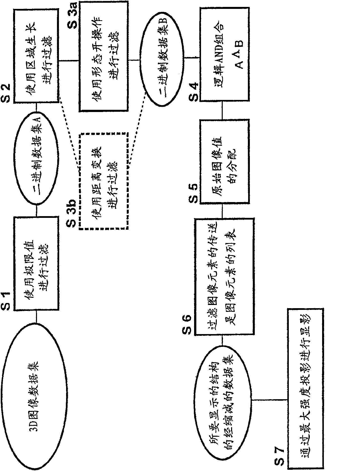
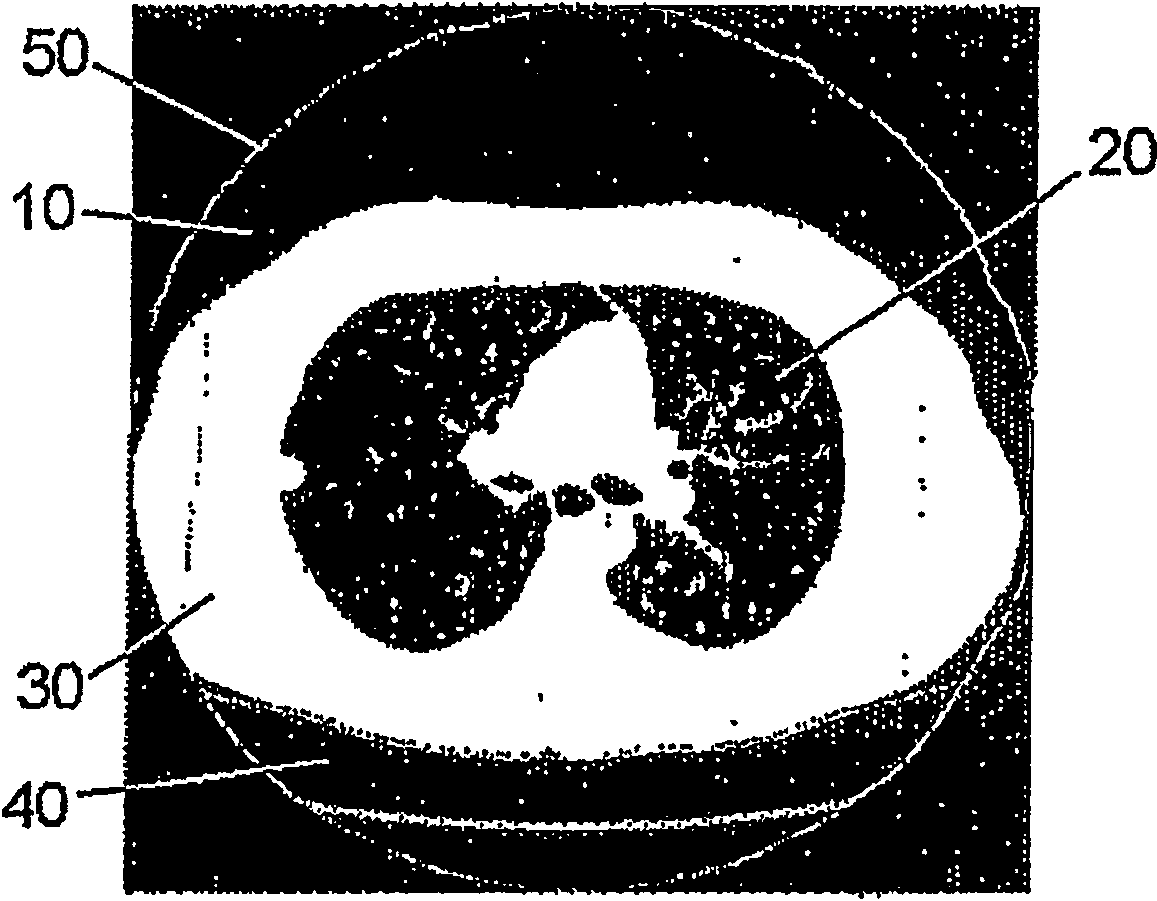
[0048] Refer to attached figure 1 , according to a preferred solution of the present invention, the input data is composed of image data of a three-dimensional dataset of the chest cavity. This data set was obtained with the aid of computed tomography. Image values ​​are associated with each image element in this 3D image dataset. This image value is usually expressed as a CT value or a so-called Hounsfield unit, which is a unit obtained by comparing the attenuation value of tissue with that of water.

[0049] The 3D image dataset is subdivided into a plurality of horizontally positioned slice images. These sliced ​​images are processed individually according to the method of the present invention.

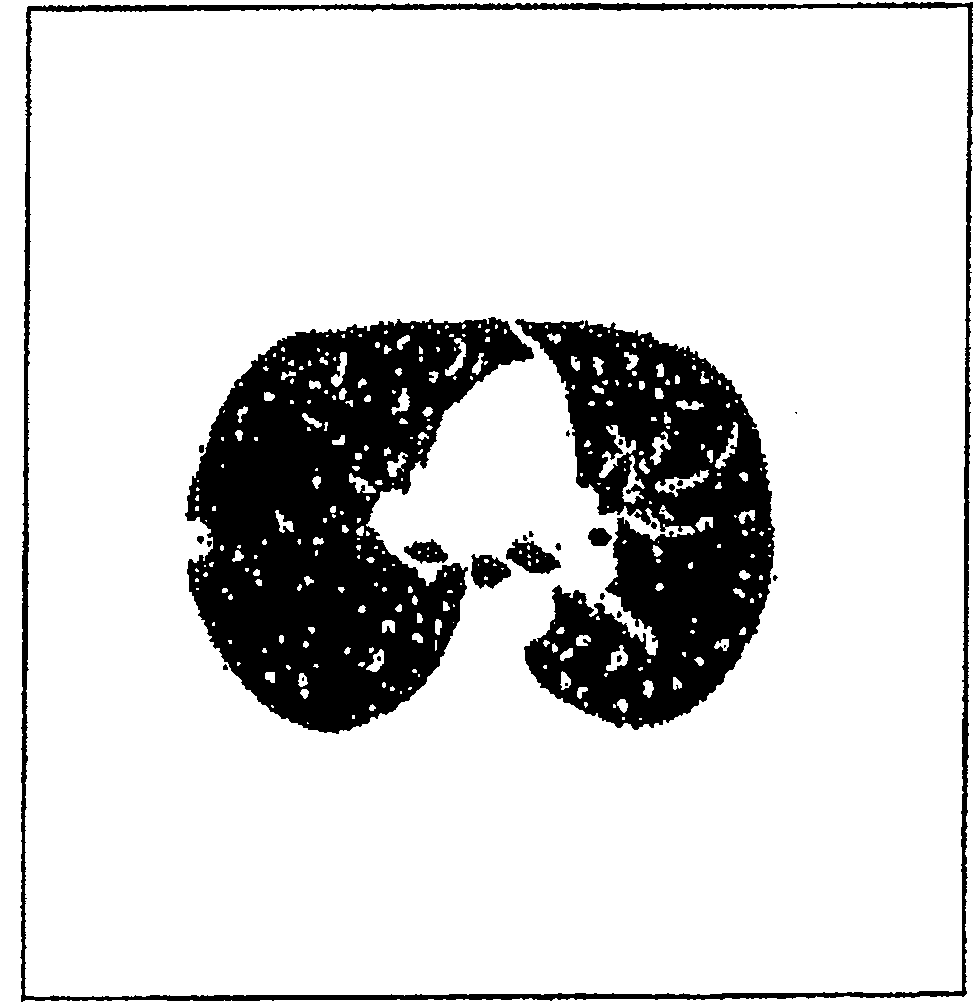
[0050] In the first step of this method (with figure 1 In S1) in the filter, a given HU is used as the limit value at this time. Set the image value of all image elements with a value HU less than this limit value (for lung tissue development, preferably equal to -400HU) to 0...


PUM


Abstract

The invention relates to a method of forming an isolated visualization of body structures from a 3D image data set, which method includes the steps of forming a binary data set in which all image elements contained in the 3D image data set are classified as image elements which are to be visualized in isolated form and image elements which are not to be visualized, where filtering of the image values is performed by means of limit values of the image values and all image elements of the binary data set of image structures contained in the 3D image data set which are smaller than a predetermined size are selected, of forming a filtered data set by entering the image values of the original 3D image data set for the image elements which are characterized as image elements to be visualized inisolated form in the binary data set, and of forming the visualization of the isolated body structure from the filtered data set. The invention also relates to a device for carrying out the describedmethod and also to programming means which carry out the described steps when executed on a computer.

Description

technical field [0001] The invention relates to a method, a device and a computer program for forming an independent visualization of human anatomy from a three-dimensional image data set. Background technique [0002] Methods of visualizing human anatomy are well known. Modern medical diagnostic technology utilizes various imaging methods to realize the functions of related examinations. Today, tomographic methods, such as computed tomography and magnetic resonance tomography, have become very important; these methods uniformly visualize the human body or its parts in the form of a plurality of discrete slice images, which are relative to each other. Positioning is performed at regular intervals. The doctor can then obtain an impression of the three-dimensional state of the structures inside the human body by observing a plurality of slice images. [0003] In slice images, human body structures having mutually different properties depending on the respective imaging meth...

Claims


Application Information

Patent Timeline
Patent Type & Authority Patents(China)
IPC IPC(8): G06T17/00G06T5/00G06T15/00A61B5/00A61B5/055G06T15/40
CPCG06T15/40
Inventor R·维克T·布拉弗特
Owner KONINK PHILIPS ELECTRONICS NV
Why Eureka
Try Eureka