Method for diagnosing testicular seminomas

A subject, selected technology, applied in the field of diagnosing testicular seminoma

Inactive Publication Date: 2005-11-30
ONCOTHERAPY SCI INC
View PDF2 Cites 8 Cited by
  • Summary
  • Abstract
  • Description
  • Claims
  • Application Information

AI Technical Summary

Problems solved by technology

[0009] Peptide-stimulated peripheral blood mononuclear cells (PBMCs) from certain healthy donors have been repeatedly reported to produce significant levels of IFN- in response to the peptide, but in 51 Cr-release assay rarely exerts cytotoxicity to tumor cells in HLA-A24 or -A0201 restricted manner (Kawano et al., Cancer Res 60:3550-8 (2000); Nishizaka et al., Cancer Res 60: 4830-7(2000); Tamura et al., Jpn J Cancer Res 92:762-7(2001))

Method used


Image

  • Method for diagnosing testicular seminomas
  • Method for diagnosing testicular seminomas
  • Method for diagnosing testicular seminomas

Examples


Embodiment 1

[0168] Example 1: Preparation of test samples

[0169] Tissues obtained from diseased tissues (for example, testicular cells from testicular germ cell tumors) and normal tissues are evaluated to identify differentially expressed genes or disease states, for example, TS. The test is carried out as follows.

[0170] Patient, tissue sample, and microdissection (LCM) obtained by laser

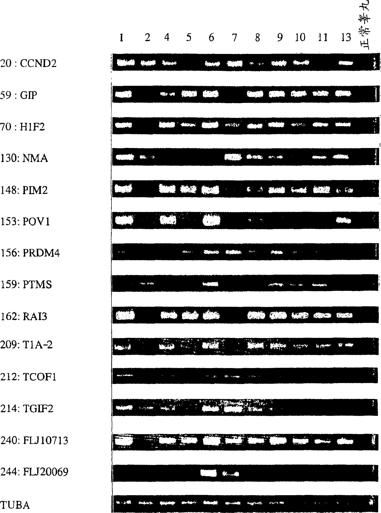
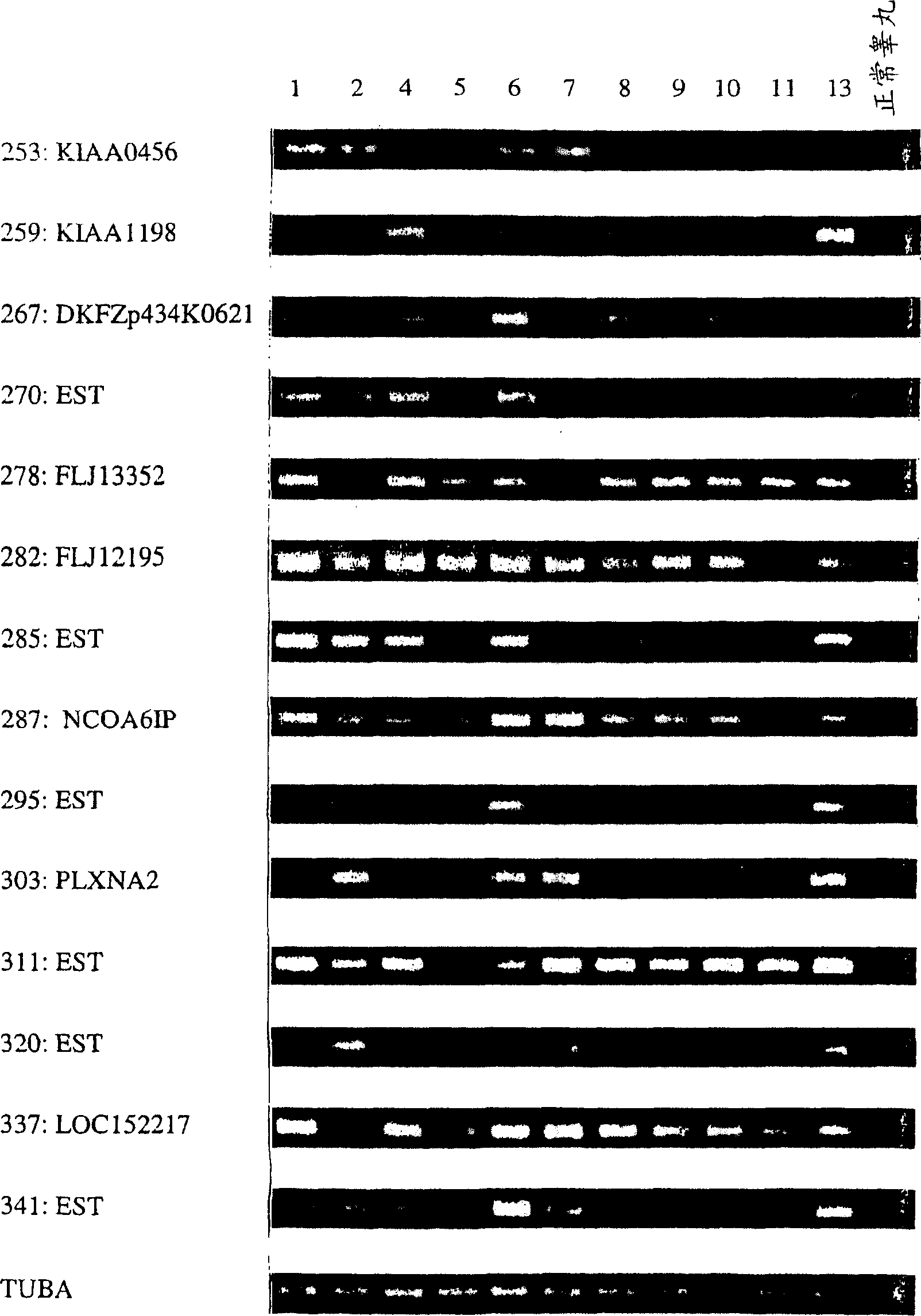
[0171] TGCT samples were obtained from 13 patients undergoing orchiectomy. The clinical characteristics of these patients are summarized in Table 1. Twelve samples diagnosed with seminoma and 1 sample with both seminoma and yolk sac tumor were used.

[0172] All samples were frozen at -80°C and then embedded in TissueTek OCT medium (Sakura). Frozen specimens were serially sectioned in 8-μm sections using a cryostat (Sakura) and stained with hematoxylin and eosin to clarify the analysis area. Then, the PixCell II LCM System (Arcturus Engineering) was used to selectively microscopically isolate seminom...

Embodiment 2

[0180] Example 2: Identification of TS-related genes

[0181]When identifying an up-regulated or down-regulated gene common to TS, analyze the gene according to the following criteria. Select the starting gene whose relative expression level can be calculated in more than 50% of cases and whose expression is up-regulated or down-regulated in more than 70% of cases. In addition, if the relative expression ratio can be calculated in 35 to 50% of cases, all cases where the gene is up- or down-regulated are also evaluated. The relative expression ratio of each gene (Cy5 / Cy3 intensity ratio) is divided into one of the following four categories: (1) Up-regulation (expression ratio exceeds 5.0); (2) Down-regulation (expression ratio less than 0.2); (3) No change in expression ( The expression ratio is between 0.2 and 5.0); and (4) No expression (or weak expression but below the detection threshold). These types are used to detect that the changes in their expression ratios are shared in ...

Embodiment 3

[0198] Example 3: Designed to reduce the growth inhibitory effect of SIRNA expressed by PYPAF3

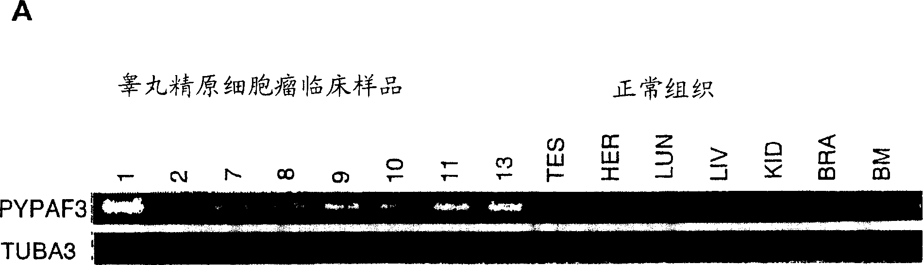
[0199] Through cDNA microarray analysis of the expression profile of the whole genome, we have isolated new molecular targets for diagnostic tumor markers, treatment and prevention of testicular germ cell tumors. Among the genes that are commonly up-regulated in testicular seminoma, we noticed that Apaf-1-like protein 3 (PYPAF3(NM 139176)) containing PYRIN was analyzed by semi-quantitative RT-PCR and included testis, heart, lung, and liver. Compared with normal human organs of kidney, brain and bone marrow, 7 out of 8 cases with testicular seminoma were significantly up-regulated. Although we currently identify PYPAF3 as an up-regulated gene in testicular seminoma (bulid#160), we initially used a cDNA microarray representing 23,040 genes retrieved from the Unigene database of the National Center for Biotechnology Information (build#131) The gene was listed as RMP: RMB5-mediated protein...


PUM


Abstract

This article describes objective methods for the detection and diagnosis of testicular seminoma (TS). In one embodiment, the diagnostic method includes determining the expression levels of TS-related genes that are differentiated between TS and normal cells. The present invention also provides methods of screening therapeutic agents for treating TS, methods of treating TS, and methods of vaccinating a subject against TS.

Description

Invention field [0001] The present invention relates to a method for diagnosing testicular seminoma. [0002] Priority information [0003] This application claims priority to the U.S. Provisional Application Serial No. 60 / 414,677 filed on September 30, 2002. technical background [0004] Although testicular germ cell tumors (TGCTs) account for 1-2% of all cancers in men, they are the most common cancers found in men aged from 20 to 40 (1) and have been onset in the past few decades The rate increased significantly (2, 3). TGCTs are divided into two main histological types, namely seminoma, which is similar to undifferentiated germ cells, and non-seminoma, which is similar to embryonic and extra-embryonic tissues, because they have either The ability of pathway differentiation (7). Seminoma is the most common histological testicular tumor among TGCTs and accounts for approximately 60% to 65% of all TGCTs (8). Currently, alpha-fetoprotein (AFP), human chorionic gonadotropin β-subu...

Claims


Application Information

Patent Timeline
Patent Type & Authority Applications(China)
IPC IPC(8): C12Q1/68
CPCC12Q1/6886C12Q2600/158C12Q2600/118C12Q2600/136A61P35/00
Inventor 中村佑辅片桐丰雅
Owner ONCOTHERAPY SCI INC
Why Eureka
Try Eureka