

Method for diagnosing testicular seminomas
- Summary
- Abstract
- Description
- Claims
- Application Information
AI Technical Summary
Benefits of technology
Problems solved by technology
Method used
Image

Examples
example 1
Preparation of Test Samples
[0155] Tissue obtained from diseased tissue (e.g., testis cells from testicular gern cell tumors) and normal tissues were evaluated to identify genes which are differently expressed or a disease state, e.g., TS. The assays were carried out as follows.
Patients, Tissue Samples and Laser-Capture Microdissection (LCM)
[0156] TGCT samples were obtained from 13 patients who underwent orchiectomy. Clinical features of these patients are summarized in Table 1. 12 samples diagnosed as seminoma and on sample of both seminoma and yolk sac tumor were used.
[0157] All samples were frozen at −80° C. and then embedded in TissueTek OCT medium (Sakura). The frozen specimens were serially sectioned in 8-μm slices with cryostat (Sakura) and were stained with hematoxylin and eosin to define the analyzed regions. Then, seminoma cells were selectively microdissected from each stained tissue with the PixCell II LCM System (Arcturus Engineering) following the manufacture's pro...
example 2
Identification of TS—Associated Genes
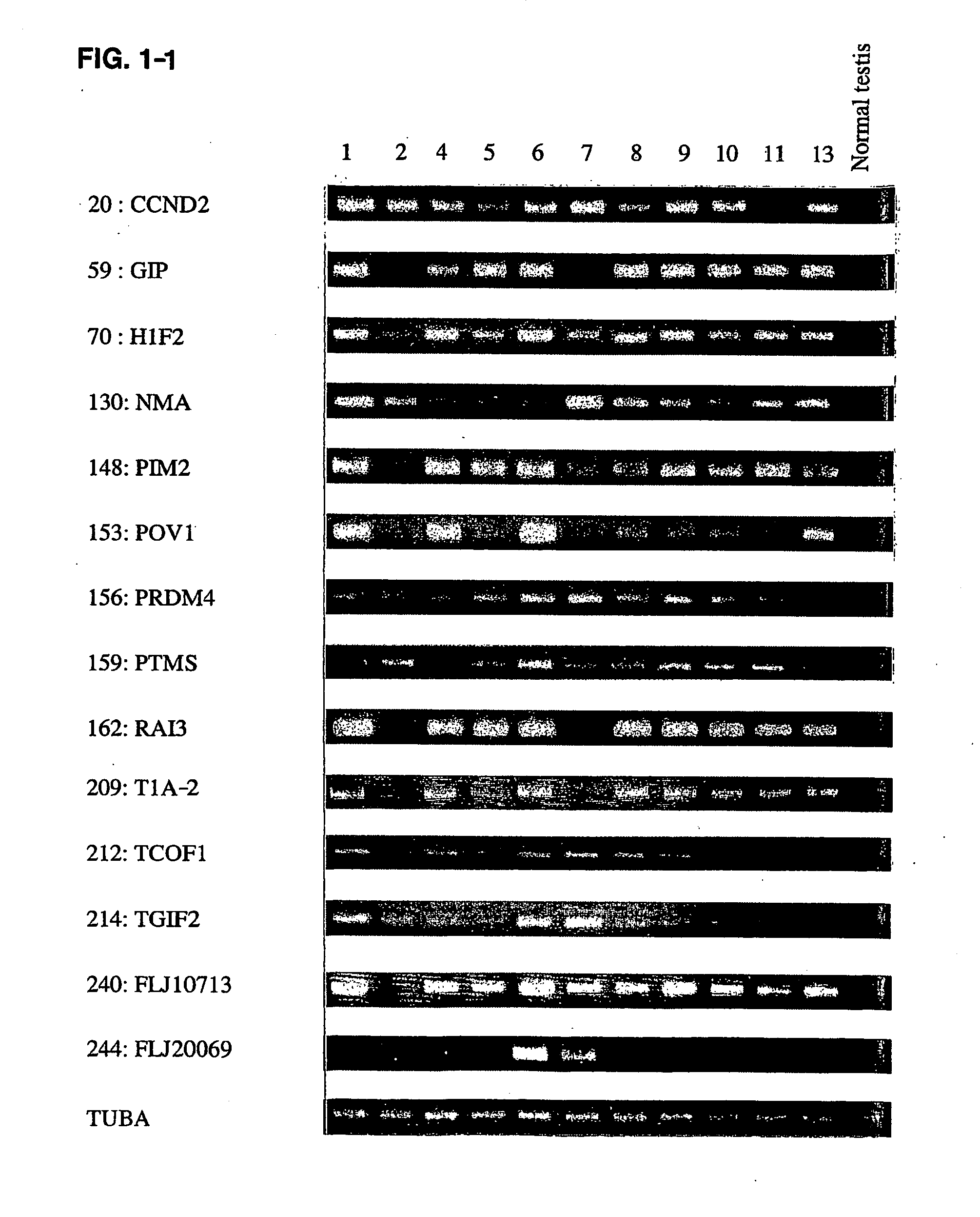
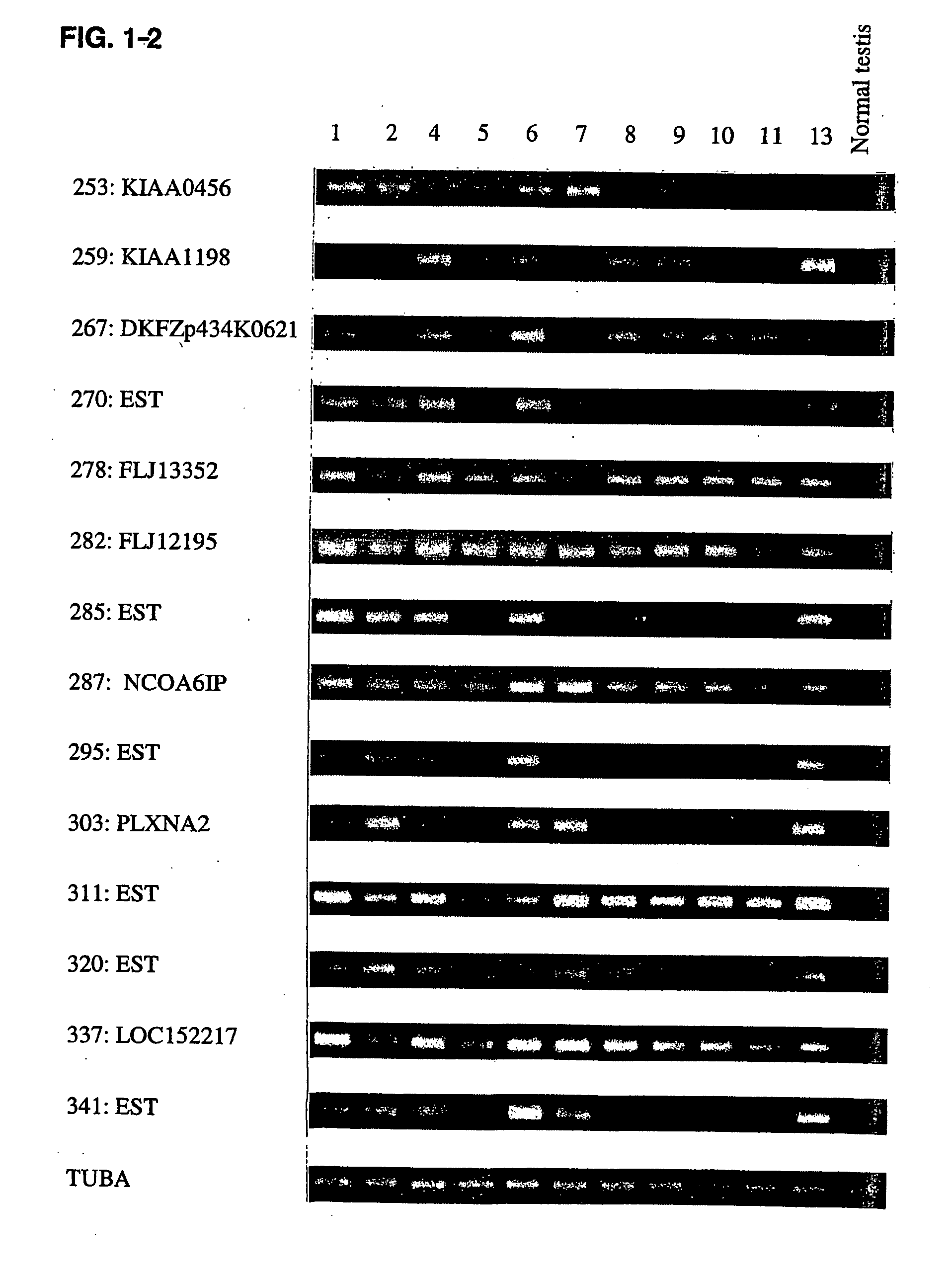
[0161] When up- or down-regulated genes common to TS were identified, the genes were analyzed according to the following criteria. Initially genes were selected whose relative expression ratio was able to calculate of more than 50% cases and whose expression were up- or down-regulated in more than 70% of cases. Moreover, if the relative expression ratio was able S to calculate of 35 to 50% cases, the genes were also evaluated that all of cases were up- or down-regulated. The relative expression ratio of each gene (Cy5 / Cy3 intensity ratio) was classified into one of four categories as follows: (1) up-regulated (expression ratio was more than 5.0); (2) down-regulated (expression ratio less than 0.2); (3) unchanged expression (expression ratio between 0.2 and 5.0); and (4) not expressed (or slight expression but under the cut-off level for detection). These categories were used to detect a set of genes whose changes in expression ratios were common...
example 3
Growth-Inhabitory Effects of siRNA Designed to Reduce Expression of PYPAF3
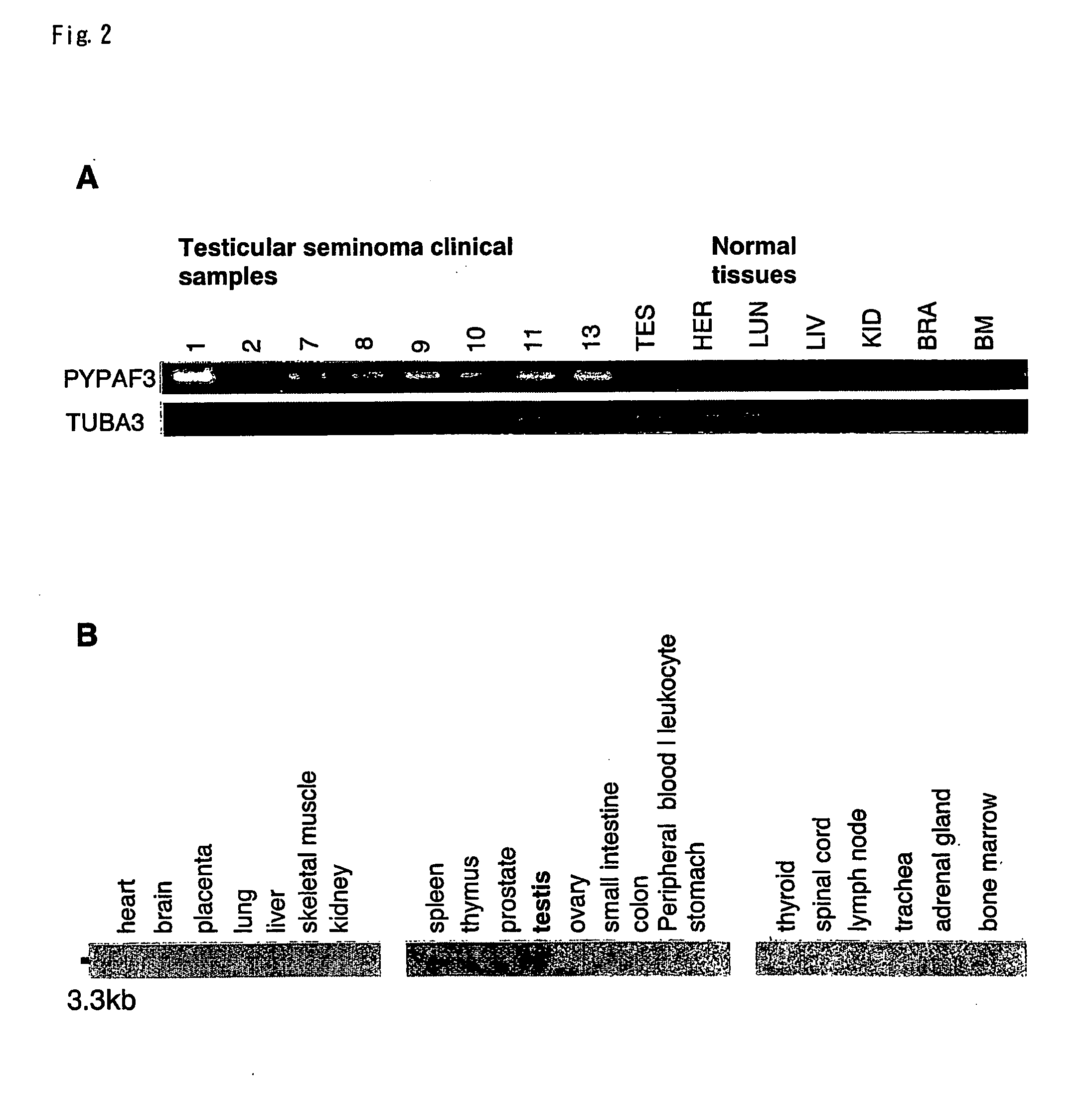
[0174] Through analysis of genome-wide expression profiles by a eDNA microarray, we have applied 5 to isolate novel molecular targets for diagnotic tumor markers, treatments and prevention of testicular germ cell tumor. Among the genes that commonly up-regulated in testicular seminomas, we focused on PYRIN-containing Apaf-1-like protein 3 (PYPAF3(NM—139176)) that were significantly up-regulated in 7 of 8 cases with testicular serninomas, compared to normal human organ including testis, heart, lung, liver, kidney, brain and bone marrow by semi-quantitative RT-PCR analysis. Although we identified PYPAF3 as up-regulated gene in testicular seminona at present (bulid #160), we initially listed this gene up as RMP:RMB5-mediating protein through expression profiles using cDNA microarray representing 23,040 genes that were retrieved from Unigene database (build #131) on Natlonal Center for Biotechnology Information. ...
PUM
| Property | Measurement | Unit |
|---|---|---|
| Fraction | aaaaa | aaaaa |
| Digital information | aaaaa | aaaaa |
| Volume | aaaaa | aaaaa |
Abstract
Description
Claims
Application Information