True three-dimensional volume imaging device with dual energy spectrum X-ray beam

An imaging device and dual-energy spectrum technology, applied in medical science, instruments for radiological diagnosis, diagnosis, etc., can solve problems such as loss of tissue structure details and easy omission of small lesions, so as to avoid loss of detailed data and improve Diagnosis effect, damage reduction effect

Inactive Publication Date: 2006-09-06
北京海思威科技有限公司
View PDF0 Cites 4 Cited by
  • Summary
  • Abstract
  • Description
  • Claims
  • Application Information

AI Technical Summary

Problems solved by technology

This CT technology uses a multi-layer spiral data acquisition and reconstruction algorithm, forming the concept of slice thickness in imaging in the direction perpendicular to the human body (defined as the Z direction), within which the human body information is averaged and lost. Therefore, the resolution in the Z direction is much smaller than that in the X-Y plane, and small lesions are easily missed.

Method used


Image

  • True three-dimensional volume imaging device with dual energy spectrum X-ray beam
  • True three-dimensional volume imaging device with dual energy spectrum X-ray beam
  • True three-dimensional volume imaging device with dual energy spectrum X-ray beam

Examples


Embodiment Construction

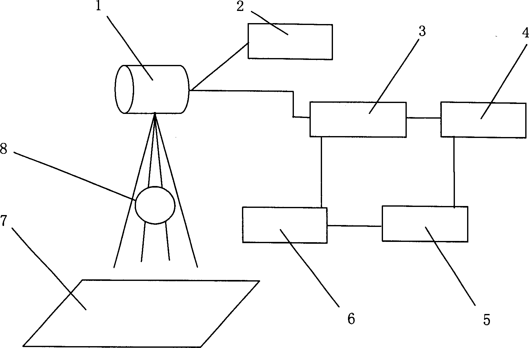
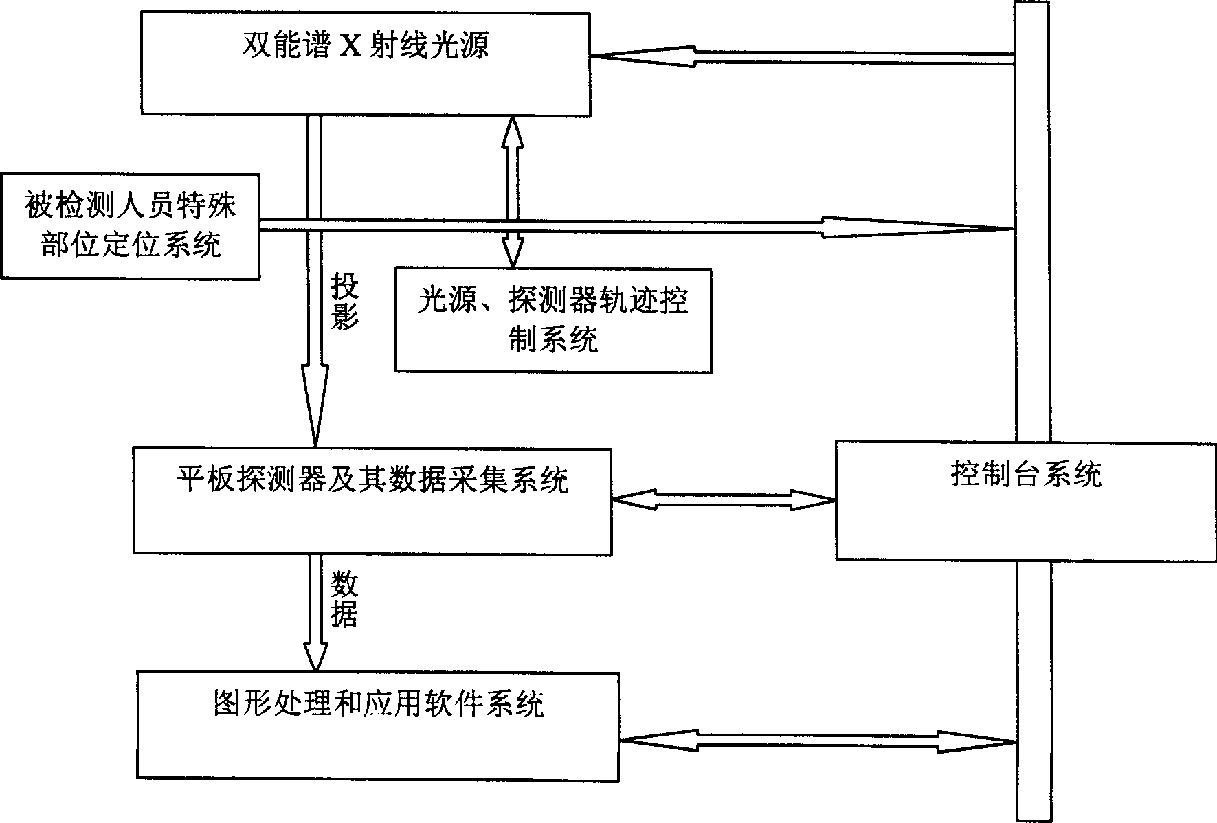
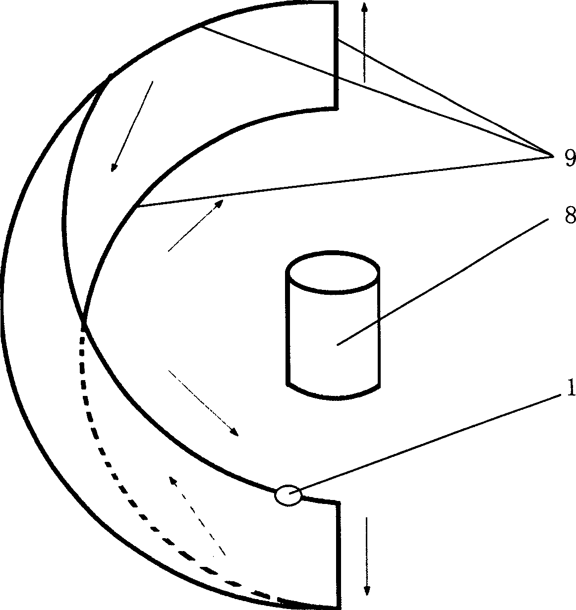
[0014] see Figure 1-Figure 5 The dual-energy spectrum true three-dimensional volume imaging device provided by the present invention includes a movable dual-energy spectrum X-ray light source 1 in the system and an X-ray flat-panel detector 7 matched therewith. In the space of the detection object 8, the beam direction of the light source faces the detection surface of the detector.

[0015] The ray beam form of the light source is preferably a cone beam, and the cone beam covers the entire object to be detected to form a projection of the object to be detected on the detector.

[0016] The light source can adopt the X-ray luminescence principle in the prior art, and is equipped with a high-voltage generator 2 and a filter collimation system. The energy spectrum width and range of the X-ray beam emitted by it can be changed according to actual needs by changing the parameters of the high-voltage generator Adjust the parameters of the filter and the filter, and the average va...


PUM


Abstract

This invention is about a dienergetic hard electron (whose energy range is 10-1400keV) three-dimension volume spectrum image-forming device, which contains removable dienergetic X-ray source orª†-radioisotope sources inside the system and the suited X-ray plate detector. The light source and the detector have a mobile linkage on a rotatable bracket, and between them there is a space for monitoring substance. The source move track can be a closed curve composed of two parallel arcs and two parallel beelines separately linking both ends of the two arcs. Its radial beam is a prick beam that always covers the whole monitoring substance, and faces to detecting side of the detector and forms monitoring substance projection on the detecting side. By using this invention, the high-resolution and isotropical three-dimension data set of soft and hard tissue on inspection personnelí»s unusual position can be obtained, and do image rebuilding image processing through computer system to form the three-dimension volume image. It can be used to physic examine flesh or inspect inner structure of industrial goods.

Description

technical field [0001] The invention relates to a digital medical diagnosis and treatment device mainly used for real three-dimensional volume imaging of human tissues. Background technique [0002] In the field of medical imaging, X-ray (including gamma-ray emitted by radioisotope) imaging device occupies a very important position. It is currently the most widely used imaging device with the fastest acquisition speed and the highest spatial resolution among all human imaging devices. , is also one of the necessary imaging devices in every hospital. Existing X-ray imaging devices are divided into two categories: X-ray plane imaging devices and tomographic three-dimensional imaging devices, among which tomographic three-dimensional imaging (CT, computed tomography) is currently the most important method for realizing three-dimensional space imaging. This CT technology uses a multi-layer spiral data acquisition and reconstruction algorithm, forming the concept of slice thickn...

Claims


Application Information

Patent Timeline
Patent Type & Authority Applications(China)
IPC IPC(8): A61B6/00A61B6/02
Inventor 包尚联贾鹏翔
Owner 北京海思威科技有限公司
Why Eureka
Try Eureka