Device and method for safely expanding minimally invasive surgical incisions

a minimally invasive, surgical technology, applied in the field of medical devices, can solve the problems of increasing the cost of the patient and the institution, low operative time with good results, and long operative time, so as to reduce the risk of accidentally cutting the recovery bag and/or the abdominal cavity, and facilitate the outward traction

Active Publication Date: 2018-08-21
UNIV OF VIRGINIA ALUMNI PATENTS FOUND
View PDF40 Cites 1 Cited by
  • Summary
  • Abstract
  • Description
  • Claims
  • Application Information

AI Technical Summary

Benefits of technology

[0008]As a brief general overview, a use by a surgeon of an embodiment of the retractor device may include, for example, when it is apparent that the gallbladder is too large to pull easily through the trocar site (e.g., cavity wall incision). Accordingly, in practice the surgeon may proceed by pushing the insertion tongue (e.g., distal portion) of the retractor device into the subject through the trocar incision, along one side of the gallbladder (typically along the side of the bag in which the gallbladder is held). The tip of the insertion tongue (distal portion) pushes the impacted gall bladder (or other organ) and its endoscopic recovery bag slightly away from the abdominal wall. A surgical tool (e.g., scalpel) is passed through the scalpel guide (aperture) with the tip of the scalpel blade in the retention groove on the back of the insertion tongue (distal portion), and with the sharp side of the scalpel blade facing away from the gallbladder and from the tongue (distal portion) of the retractor device. The scalpel is then passed down through the abdominal wall, with its tip in the retention groove, while tension on the retractor device handle away from the gallbladder, and gentle outward traction on the gallbladder (and bag) permits removal of the gallbladder (and bag) as soon as the opening is just large enough to allow it. This step may be repeated if the gallbladder cannot be removed after a single passage of the scalpel. The retractor device also may be removed before the gallbladder and its bag are pulled through the enlarged incision. When properly used, the design of the retractor device mitigates the risk of accidentally cutting the recovery bag and / or organ as the incision is enlarged, and also keeps the opening occluded to prevent decompression of the abdominal cavity, thus keeping the abdominal wall and the scalpel blade used to extend the incision well away from abdominal contents.

Problems solved by technology

Operative times are low with good outcomes; however, one source of frustration commonly encountered involves extraction of the gallbladder from the patient's abdomen through an incision often smaller than the gallbladder itself.
However, in difficult cases the awkward maneuvers in conjunction with repeat attempts can lead to lengthened operative times, with increased cost to the patient and the institution.
This difficult problem may add 20 minutes or more to a procedure which otherwise can take as little as one hour.
Operating room time is expensive; current estimates of cost is about $30 per minute, thus saving 20 minutes could save over $600.
Bile that spills into the abdomen is irritating and can cause inflammatory changes and result in infection.
Bile spillage is particularly undesirable in patients harboring a gallbladder cancer because of concern for port site or peritoneal metastasis.
Few studies have attempted to discern the optimal method for gallbladder extraction from the abdominal cavity; however, one study found incision extension to result in no increase in morbidity.
Unfortunately, such extension is often technically awkward and entails significant risk to the patient, because available tools are not designed for this purpose other than the endoscopic bags that are used to contain the tissue as it is pulled through the abdominal wall.
Collectively, these approaches can result in sequelae of stretching such as bruising and excessive pain, or may result in puncture of the specimen bag, which should be avoided given the associated risks mentioned above.

Method used


Image

  • Device and method for safely expanding minimally invasive surgical incisions
  • Device and method for safely expanding minimally invasive surgical incisions
  • Device and method for safely expanding minimally invasive surgical incisions

Examples


examples

[0064]Practice of an aspect of an embodiment (or embodiments) of the invention will be still more fully understood from the following example set, which is presented herein for illustration only and should not be construed as limiting the invention in any way.

examples set no.1

Examples Set No. 1

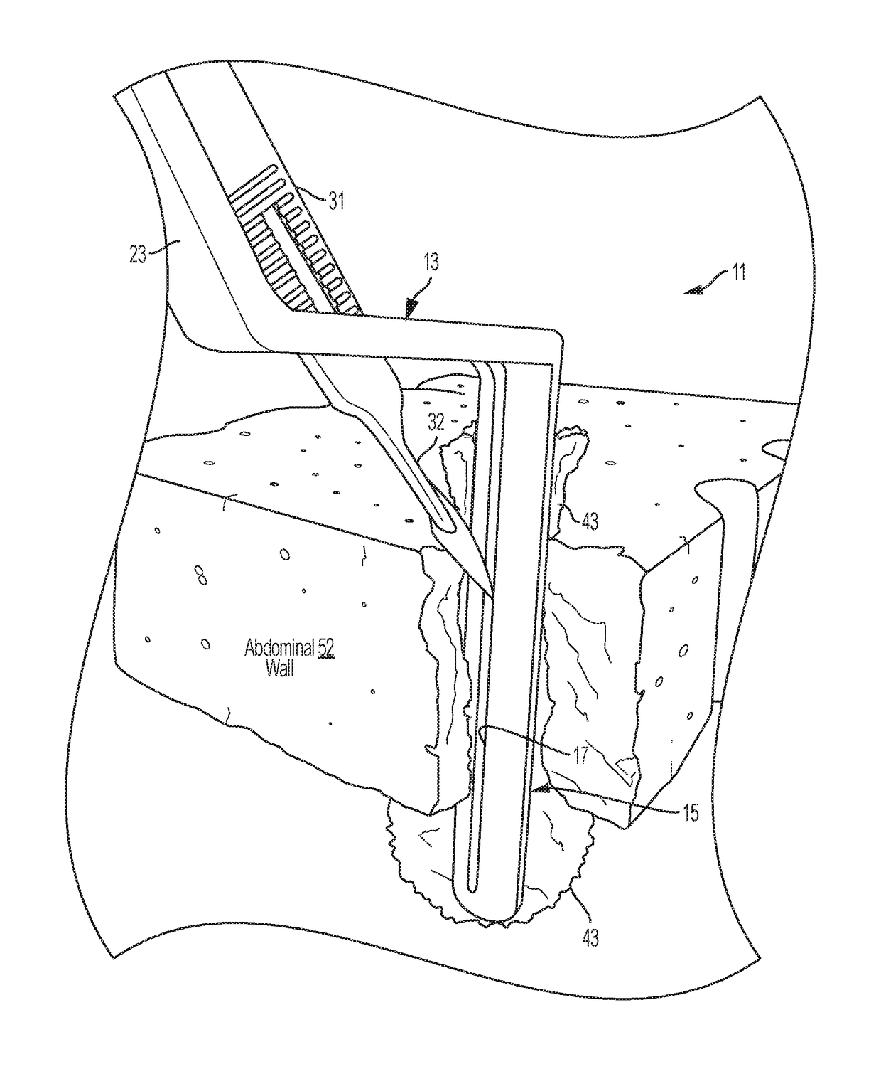
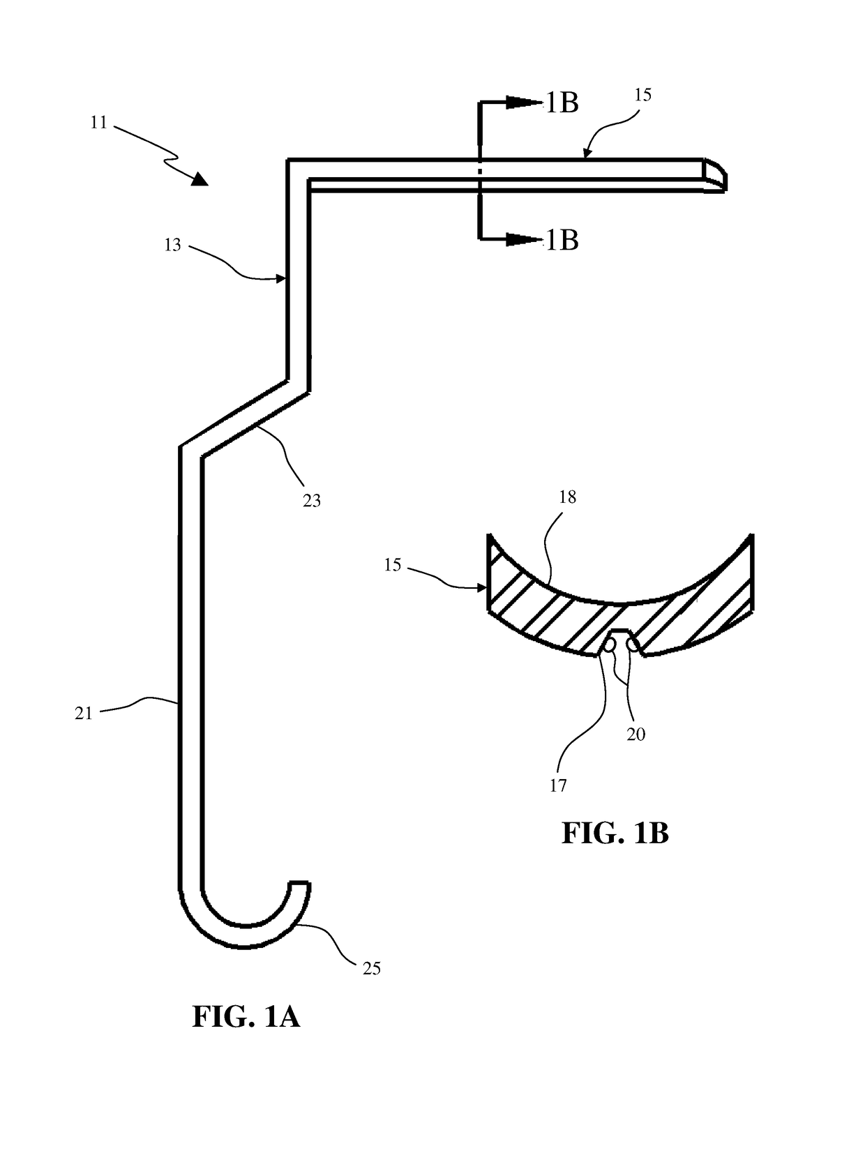
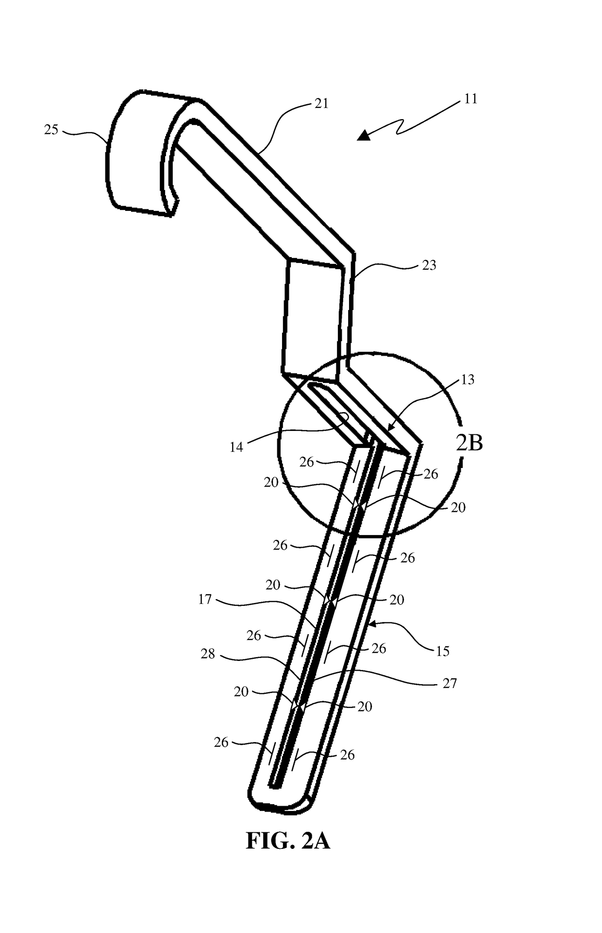
[0065]For instance, in a prototype a single-piece stainless steel surgical instrument, the purpose of which is to help remove impacted endoscopic recovery bags and organs and eliminate the risk of damaging the organ. An aspect of the prototype provides, a single-piece stainless steel surgical instrument consisting of a handle designed to be gripped in a single hand. The handle includes a curved end (catch) to provide an easy grip or leverage for a surgeon to hold. The handle is attached to a flat extension region (i.e., portion of a proximal portion) with a slot (or aperture) just wide enough to accommodate the width of a standard surgical scalpel handle (thereby providing a scalpel slot). The scalpel slot (aperture) is oriented along the midline of the flat extension region (i.e., portion of the proximal portion), beginning where the extension region meets the handle to the extreme end of the flat region opposite the handle, at the junction with a tongue (i.e., di...


PUM


Abstract

An insertion tongue of the retractor device may be pushed into the subject through the trocar incision, along one side of the organ. The tip of the insertion tongue pushes the impacted organ and its endoscopic recovery bag slightly away from the cavity wall. A scalpel is passed through the insertion guide with the tip of the scalpel blade in the retention guide on the back of the insertion tongue, and with the sharp side of the scalpel blade facing away from the organ and from the tongue of the retractor device. The scalpel is then passed down through the cavity wall, with its tip in the retention guide, while tension on the retractor device handle away from the organ, and gentle outward traction on the organ (and bag) permits removal of the organ (and bag) as soon as the opening is just large enough to allow it.

Description

RELATED APPLICATIONS[0001]This application is a national stage filing of International Application No. PCT / US2011 / 059379, filed Nov. 4, 2011, which claims the benefit of priority under 35 U.S.C. Section 119(e) from U.S. Provisional Application Ser. No. 61 / 410,040 filed Nov. 4, 2011, entitled “Laparo-Extender Device and Method for Safely Expanding Laparoscopic Surgical Incisions;” the disclosures of which are hereby incorporated by reference herein in their entirety.FIELD OF THE INVENTION[0002]The present invention relates generally to the field of medical devices to be used for minimally invasive procedures. More specifically, the invention is in the subfield of retractor devices and other related tools to be used for surgical procedure types such as laparoscopic, thoracic, or endoscopic.BACKGROUND OF THE INVENTION[0003]With an estimated 500,000 minimally invasive cholecystectomies performed each year, laparoscopic cholecystectomy is one of the most common general surgical procedure...

Claims


Application Information

Patent Timeline
Patent Type & Authority Patents(United States)
IPC IPC(8): A61B17/02A61B17/32
CPCA61B17/02A61B2017/320052
Inventor GUILFORD, WILLIAM H.SLINGLUFF, JR., CRAIG L.JUDGE, JOSHUA M.
Owner UNIV OF VIRGINIA ALUMNI PATENTS FOUND
Why Eureka
Try Eureka