
Mechanical stress response analysis of cells and tissues

a cell and tissue technology, applied in the field of mechanical stress response analysis of cells and tissues, can solve the problems of low throughput, time-consuming and difficult to analyze, and use a very limited number of cells per day

Active Publication Date: 2018-12-18
RGT UNIV OF CALIFORNIA
View PDF6 Cites 0 Cited by
  • Summary
  • Abstract
  • Description
  • Claims
  • Application Information

AI Technical Summary

Benefits of technology

The patent text describes a method and device for discovering and screening compounds that can modulate cellular mechanotransduction. The method involves administering a test compound to a cellular sample and initiating a microcavitation bubble to monitor the cellular response. A difference in the signal compared to a control indicates that the test compound has modulated cellular mechanotransduction. The device includes a holder for holding cellular samples and an energy source for generating a microcavitation bubble. The technical effect of this patent is a high-throughput tool for identifying and testing compounds that can affect how cells respond to mechanical stimuli.

Problems solved by technology

While these methods are well-suited for applying physiological forces to cells in two-dimensional (2-D) and three-dimensional (3-D) cell cultures, they are time-intensive and require considerable technical expertise.
Some of these methods, including optical tweezers and AFM, have utility in the study of mechanotransduction, but their low throughput means that they can only be used to examine a very limited number of cells per day.
While such systems are well-suited to screen a small number of molecules shown to affect mechanotransduction, there is no clear path to scaling up such systems for screening hundreds of thousands of molecules.
As described above, current methods are not standardized and they are not compatible with existing high-throughput drug discovery platforms.
Thus, the technical challenge involved in developing a high-throughput platform for applying precise forces to multiple cells in various culture conditions is considerable.

Method used


Image

  • Mechanical stress response analysis of cells and tissues
  • Mechanical stress response analysis of cells and tissues
  • Mechanical stress response analysis of cells and tissues

Examples


examples

Laser Microscope

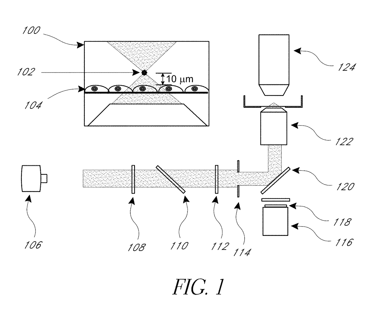
[0082]μCBs were created using the λ=532 nm emission of a Q-switched pulsed microchip laser (PNG-M03012, Teem Photonics) emitting pulses 500 ps in duration. The laser beam is expanded and collimated and the pulse energy of the collimated beam is controlled using the combination of a λ / 2 waveplate and polarizing beam splitter. The central portion of the beam is selected by an iris and directed into an inverted microscope (Olympus IX-81) by a dichroic (Chroma ZT532NBDC) mirror. The beam is then focused by a 20×, 0.45 numerical aperture microscope objective (Olympus IX-81). The laser microbeam was focused approximately 10 μm above the cell monolayer.

[0083]Fluorescence microscopy was performed on the same inverted microscope using epifluorescence illumination from a mercury short-arc lamp (X-Cite 120PC, Lumen Dynamics). The filter cube containing a 480 / 40 excitation, 535 / 50 emission, and 505 LP dichroic filters (Chroma) which were chosen based on the fluorescent probe spe...


PUM

PropertyMeasurementUnit
diameteraaaaaaaaaa
diameteraaaaaaaaaa
diameteraaaaaaaaaa

Abstract

A method and device are disclosed for inducing mechanical stress in a cellular sample to evaluate mechanotransduction in the cellular sample. In one embodiment, the mechanical stress is induced by generating a microcavitation bubble in the cellular sample using a pulsed energy. The microcavitation bubble creates a microtsunami, which provides a transient, impulsive mechanical stress on the cellular sample, forming a gradient of effects at distances away from the microcavitation bubble.

Description

STATEMENT REGARDING FEDERALLY SPONSORED R&D[0001]This research was funded through the Laser Microbeam and Medical Program, a National Biomedical Technology Resource (P41-EB015890) supported by the National Institutes of Health.INCORPORATION BY REFERENCE TO ANY PRIORITY APPLICATIONS[0002]Any and all applications for which a foreign or domestic priority claim is identified in the Application Data Sheet as filed with the present application are hereby incorporated by reference under 37 CFR 1.57.BACKGROUND OF THE INVENTION[0003]Field of the Invention[0004]Methods are disclosed for screening mechanical stress responses in cells and tissues using induced cavitation bubbles.[0005]Description of the Related Art[0006]Mechanotransduction, which refers to the mechanical forces resulting from cell-cell and cell-matrix interactions, are known to influence the signaling, function, homeostasis, and fate of individual cells and cell populations. Likewise, numerous studies have demonstrated the impo...

Claims


Application Information

Patent Timeline
Patent Type & Authority Patents(United States)
IPC IPC(8): G01N33/50C40B30/06G01N33/543
CPCG01N33/5032G01N33/54373G01N33/5064C40B30/06
Inventor BOTVINICK, ELLIOT L.VENUGOPALAN, VASANCOMPTON, JONATHANHELLMAN, AMY
Owner RGT UNIV OF CALIFORNIA
Learn More