Medical imaging apparatus having multiple subsystems, and operating method therefor

a medical imaging and subsystem technology, applied in the direction of instruments, reradiation, measurement using nmr, etc., can solve the problems of complex systems of magnetic resonance apparatuses and computed tomography apparatuses, difficult for users to combine with other users, and worsen image quality

Active Publication Date: 2019-01-29
SIEMENS HEALTHCARE GMBH
View PDF11 Cites 0 Cited by
  • Summary
  • Abstract
  • Description
  • Claims
  • Application Information

AI Technical Summary

Benefits of technology

[0011]An object of the present invention is to provide a method for the operation of a medical imaging examination apparatus that has multiple subsystems, and a control computer that controls the subsystems in a coordinated manner in order to carry out a scan sequence, a medical imaging examination apparatus and an associated electronically readable data carrier, which enable reliable adaptation of sequence control data to existing ambient conditions (adjustment) in a user-friendly and time-optimized manner and overcomes the above-mentioned disadvantages of conventional methods.
[0014]With the method according to the invention and the central determination therein of control signals even before the actual scan, it is made possible to use a dynamic adjustment and thus optimization of control signals reliably, in a user-friendly way and time-optimized, including in routine clinical practice. Herein, restriction in a targeted manner to subregions in a volume of interest of the examination object is possible, and this has a positive effect on the computation time and on the quality of the examination results. By the restriction to respective relevant sequence control data and the determination and making available of the optimized control signals, the data flow is optimized and therefore can be easily integrated into existing workflows. By the improved possibilities for adapting the control signals, it is also possible to build less expensive hardware into the medical imaging examination apparatuses used, and thereby to manufacture these devices more economically, since any sub-optimum ambient conditions due to the hardware can be compensated for with the method. Thus, with the method according to the invention, an optimized integration of the scans, evaluations and uses employed for determining the optimized control signals into the technical sequence of the scan of the data from the examination object through the control protocol is provided.
[0015]In the inventive method, it is not necessary, as in the prior art, for a user to ensure the consistency of different scan protocols, for example, protocols for adjustment scans for determining ambient conditions and from the actual scan. Rather, established adjustment scans can be used without a user having to parameterize them first.
[0016]By the determination of control signals according to the invention for different states of physiological processes occurring, a consistent quality of the scan data can be achieved even under temporal variation of the ambient conditions depending on the physiological processes. Herein, at most a slight intervention into the ongoing scan is necessary in order to determine current states of the physiological processes.

Problems solved by technology

Medical imaging examination apparatuses such as magnetic resonance apparatuses and computed tomography apparatuses are complex systems with a large number of technical subsystems.
The user needs to watch very closely for consistency between the imaging sequence and the field determination sequence since, otherwise, inconsistencies can lead to a worsening of the image quality.
These methods are therefore very complex and difficult for the user to combine with other, for example static, adjustments since interactions between different parameters cannot be taken into account or only to a limited extent.
Furthermore, the optimization is restricted to the slices of the imaging sequence.
Due to the determination of the parameters at run time, it can however occur that, in the time available which is limited due to the already running scan sequence, no useful parameters can be determined.

Method used


Image

  • Medical imaging apparatus having multiple subsystems, and operating method therefor
  • Medical imaging apparatus having multiple subsystems, and operating method therefor
  • Medical imaging apparatus having multiple subsystems, and operating method therefor

Examples


Embodiment Construction

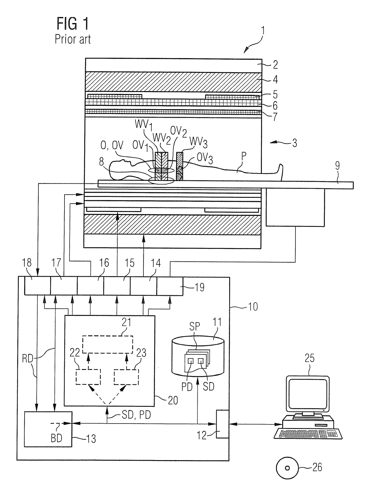
[0025]FIG. 1 shows a basic schematic form of a medical imaging examination apparatus 1 that although the basic components are known, can be configured according to the invention. The apparatus includes the actual magnetic resonance scanner 2 with an examination space 3 or patient tunnel situated therein. A table 9 can be moved into this patient tunnel 3 through various positions so that an examination object, e.g. a patient P or test subject lying thereon can be placed during an examination at a particular position within the magnetic resonance scanner 2 relative to the magnetic system and the radio frequency system arranged therein and is also displaceable between different positions during a scan. It should be mentioned at this point that the exact construction of the magnetic resonance scanner 2 is not essential. Thus, for example, a cylindrical system with a typical patient tunnel can be used, but also a C-arm-shaped magnetic resonance device which is open at one side.

[0026]Basi...


PUM


Abstract

In a method for operating a medical imaging apparatus having subsystems, a control protocol assigned to a scan sequence to be performed is provided to a control computer that determines sequence control data for the control protocol, which define different functional subsequences of the scan sequence. Different effective volumes are assigned to each functional subsequence, and current ambient conditions of the apparatus are determined for the sequence control data and associated effective volumes, for a series of states of physiological processes that occur during the scan sequence. Control signals for the scan sequence are determined from the sequence control data, the effective volumes and the current ambient conditions per observed state, that optimize the functional subsequences of the scan sequence locally. During the scan, at least one current state of the observed physiological processes is determined and the control signals used for the ongoing scan are determined from the stored control signals, dependent on the at least one current state.

Description

BACKGROUND OF THE INVENTIONField of the Invention[0001]The invention concerns to the operation of a medical imaging examination apparatus having multiple subsystems, a corresponding medical imaging examination apparatus, and an associated electronically readable data carrier encoded with programming instructions for implementing such a method.Description of the Prior Art[0002]Medical imaging examination apparatuses such as magnetic resonance apparatuses and computed tomography apparatuses are complex systems with a large number of technical subsystems. These include, in a magnetic resonance apparatus, a basic field magnet system, a gradient system, a shim system and a radio frequency transmission system as well as a radio frequency receiving system.[0003]In order to generate images or spectroscopic data from an examination object with a magnetic resonance apparatus, the examination object is positioned in the scanner in a strong homogeneous basic magnetic field, also known as the B0...

Claims


Application Information

Patent Timeline
Patent Type & Authority Patents(United States)
IPC IPC(8): G01V3/00A61B5/055G01R33/54G01R33/483
CPCG01R33/543A61B5/055G01R33/4835
Inventor BENNER, THOMASCAMPAGNA, SWENFEIWEIER, THORSTENKUEHN, BERNDSPEIER, PETER
Owner SIEMENS HEALTHCARE GMBH
Why Eureka
Try Eureka