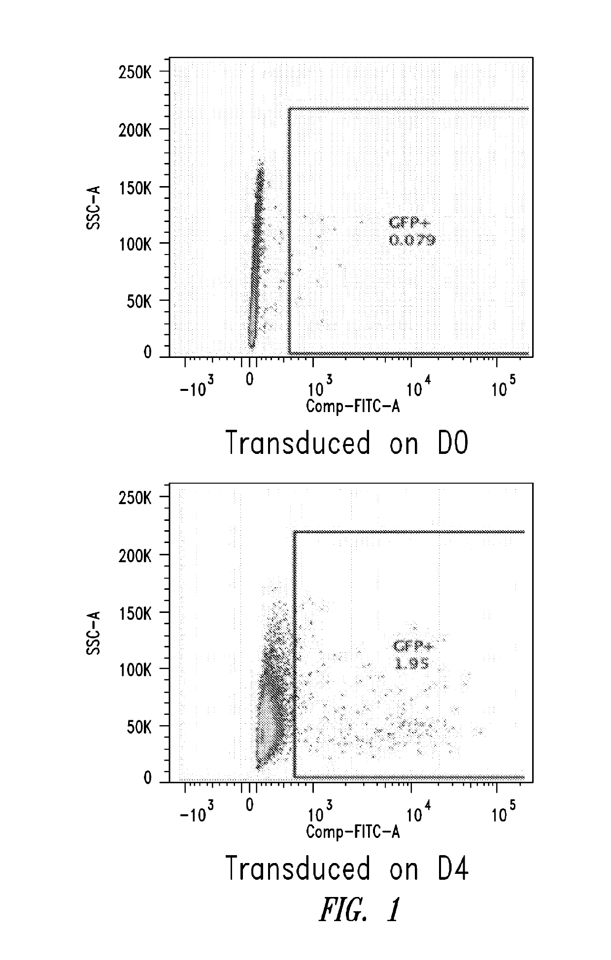
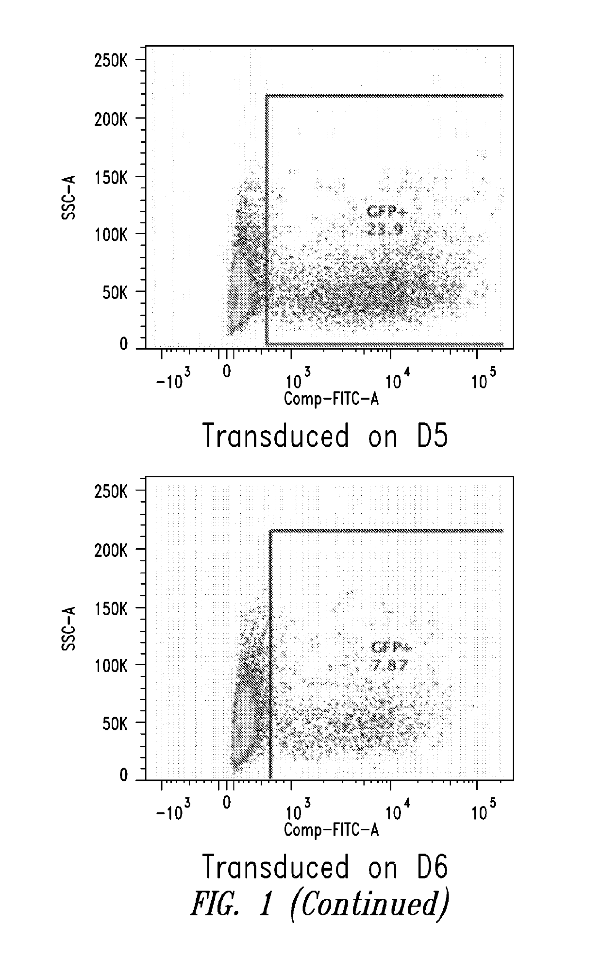
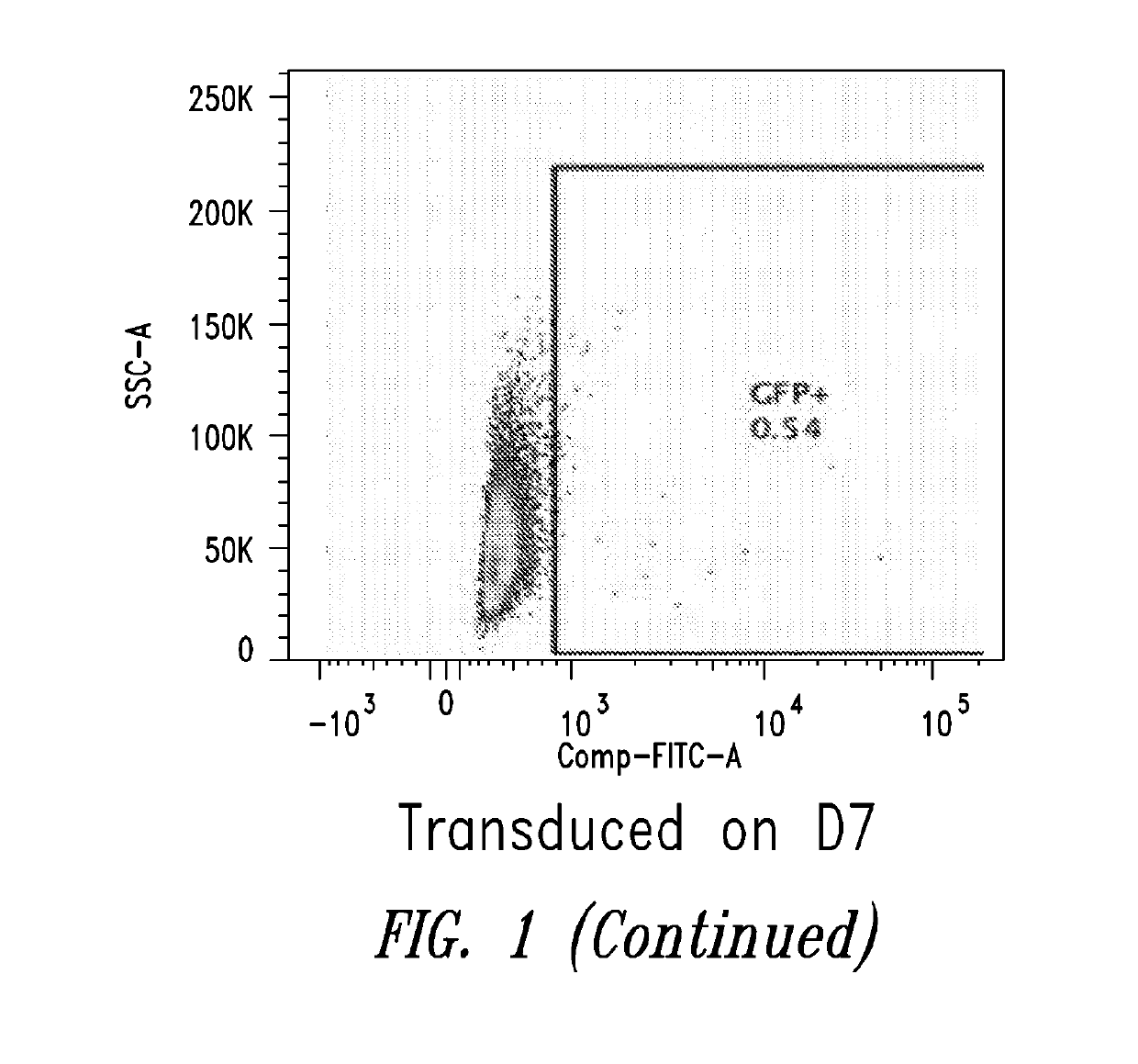
[0014]The present disclosure relates generally to culturing B cells in vitro under conditions so as to differentiate memory B cells into plasmablasts and plasma cells. The present invention further relates to genetically modifying B cells to express a gene of interest and activating and culturing the modified B cells so as to express the protein encoded by the gene of interest. These activated modified B cells are then administered to a subject. The compositions and methods described herein provide the ability to differentiate in vitro B cells derived from, for example, a simple blood draw, and then transduce the differentiated B cells with a nucleic acid of interest such that they secrete abundant amounts of the protein encoded by the nucleic acid of interest. One particular advantage of this system is that it only requires about 10 days to differentiate a resting memory B cell into a plasmablast or plasma cell, which can then be used for transduction and production of a protein of interest. Systems known in the art can take as long as 10 weeks. Thus, the present disclosure provides methods that require less time in culture providing commercial and safety advantages as compared to prior methods. An additional advantage of the present invention is that the in vitro culture methods provide increased transduction efficiency to about 20% transduction efficiency. This is an improvement over the low transduction efficiencies of methods known in the art. The present methods also do not require feeder cells which are burdensome for GMP grade production.
[0015]The prototypic example of cells for use with the transduction methods of the disclosure is B cells. In certain embodiments, the B cells to be used in the methods of this disclosure may be resting B cells, memory B cells, primed or activated B cells, myeloma cells, lymphocytic leukemia B cells, or B lymphoma cells. However, one of ordinary skill in the art will readily appreciate that the vectors and methods of the disclosure may be applied to other cell types. By way of example, cell types that may be cultured, differentiated, modified and activated include any typically nondividing or quiescent cell such as neuroblasts, hematopoietic stem cells and hematopoietic progenitor cells (CD34+ cells), mesenchymal stem cells, mesenchymal progenitor cells, neural and hepatic progenitor and stem cells, dendritic cells, T cells (CD8+ or CD4+ T cells), other leukocyte populations, pluripotent stem cells, multi-potent stem cells, etc. Accordingly, certain embodiments of the present disclosure provides populations of cells resulting from this methodology. In certain embodiments, the methods and vectors as described herein may be used to transduce any other cell type desired, such as hepatocytes, epithelial cells, osteoblasts, myocytes, fibroblasts, adipocytes etc.
[0016]“Quiescent”, as used herein, refers to a cell state wherein the cell is not actively proliferating.
[0017]Prior to differentiation and transduction, a source of B cells is obtained from a subject. The term “subject” is intended to include living organisms in which an adaptive immune response can be elicited (e.g., mammals). Examples of subjects include humans, dogs, cats, mice, rats, and transgenic species thereof. B cells can be obtained from a number of sources, including peripheral blood mononuclear cells, bone marrow, lymph node tissue, cord blood, tissue from a site of infection, spleen tissue, and tumors. In certain embodiments of the present disclosure, any number of B cell lines available in the art, may be used. In certain embodiments of the methods described herein, B cells can be obtained from a unit of blood collected from a subject using any number of techniques known to the skilled artisan, such as FICOLL™ (copolymers of sucrose and epichlorohydrin that may be used to prepare high density solutions) separation. In one preferred embodiment, cells from the circulating blood of an individual are obtained by apheresis or leukapheresis. The apheresis product typically contains lymphocytes, including T cells, monocytes, granulocytes, B cells, other nucleated white blood cells, red blood cells, and platelets. In one embodiment, the cells collected by apheresis may be washed to remove the plasma fraction and to place the cells in an appropriate buffer or media for subsequent processing steps. In one embodiment of the methods described herein, the cells are washed with phosphate buffered saline (PBS). In an alternative embodiment, the wash solution lacks calcium and may lack magnesium or may lack many if not all divalent cations. As those of ordinary skill in the art would readily appreciate a washing step may be accomplished by methods known to those in the art, such as by using a semi-automated “flow-through” centrifuge (for example, the Cobe 2991 cell processor) according to the manufacturer's instructions. After washing, the cells may be resuspended in a variety of biocompatible buffers, such as, for example, PBS. Alternatively, the undesirable components of the apheresis sample may be removed and the cells directly resuspended in culture media.
[0018]B cells may be isolated from peripheral blood or leukapheresis using techniques known in the art. For example, PBMCs may be isolated using FICOLL™ (Sigma-Aldrich, St Louis, Mo.) and CD19+ B cells purified by negative or positive selection using any of a variety of antibodies known in the art, such as the Rosette tetrameric complex system (StemCell Technologies, Vancouver, Canada). In certain embodiments, memory B cells are isolated as described by Jourdan et al., (Blood. 2009 Dec. 10; 114(25):5173-81). For example, after removal of CD2+ cells using anti-CD2 magnetic beads, CD19+ CD27+ memory B cells can be sorted by FACS. Bone marrow plasma cells (BMPCs) can be purified using anti-CD138 magnetic microbeads sorting or other similar methods and reagents.
[0019]Other isolation kits are commercially available, such as R&D Systems' MagCellect Human B Cell Isolation Kit (Minneapolis, Minn.). In certain embodiments, resting B cells may be prepared by sedimentation on discontinuous Percoll gradients, as described in (Defranco et al., (1982) J. Exp. Med. 155:1523).