Systems and methods for camera-based image processing in microscopy instruments

Active Publication Date: 2019-03-26
LEICA MICROSYSTEMS CMS GMBH
View PDF9 Cites 1 Cited by
  • Summary
  • Abstract
  • Description
  • Claims
  • Application Information

AI Technical Summary

Benefits of technology

The patent describes a system and method for taking high-resolution pictures of a specimen using a fluorescence microscope. The system includes a camera that can control the output of its bright light and capture images of the specimen. These images are then processed using special techniques to create a final, high-resolution image of the specimen. Overall, this technology allows for better and more precise observation of small samples.

Problems solved by technology

While acquisition of the images can be accomplished relative quickly, handling and processing such a large number of images is relatively slow because these techniques necessitate acquiring and processing a large amount of image data in order to obtain a relatively small number of pixel locations.

Method used


Image

  • Systems and methods for camera-based image processing in microscopy instruments
  • Systems and methods for camera-based image processing in microscopy instruments
  • Systems and methods for camera-based image processing in microscopy instruments

Examples


Embodiment Construction

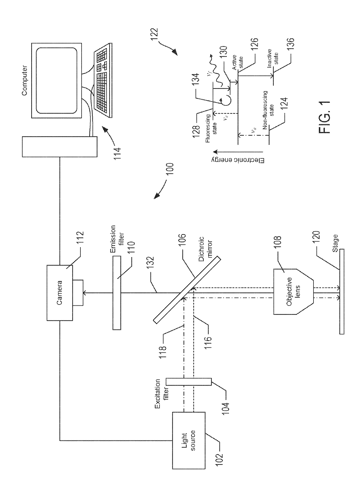
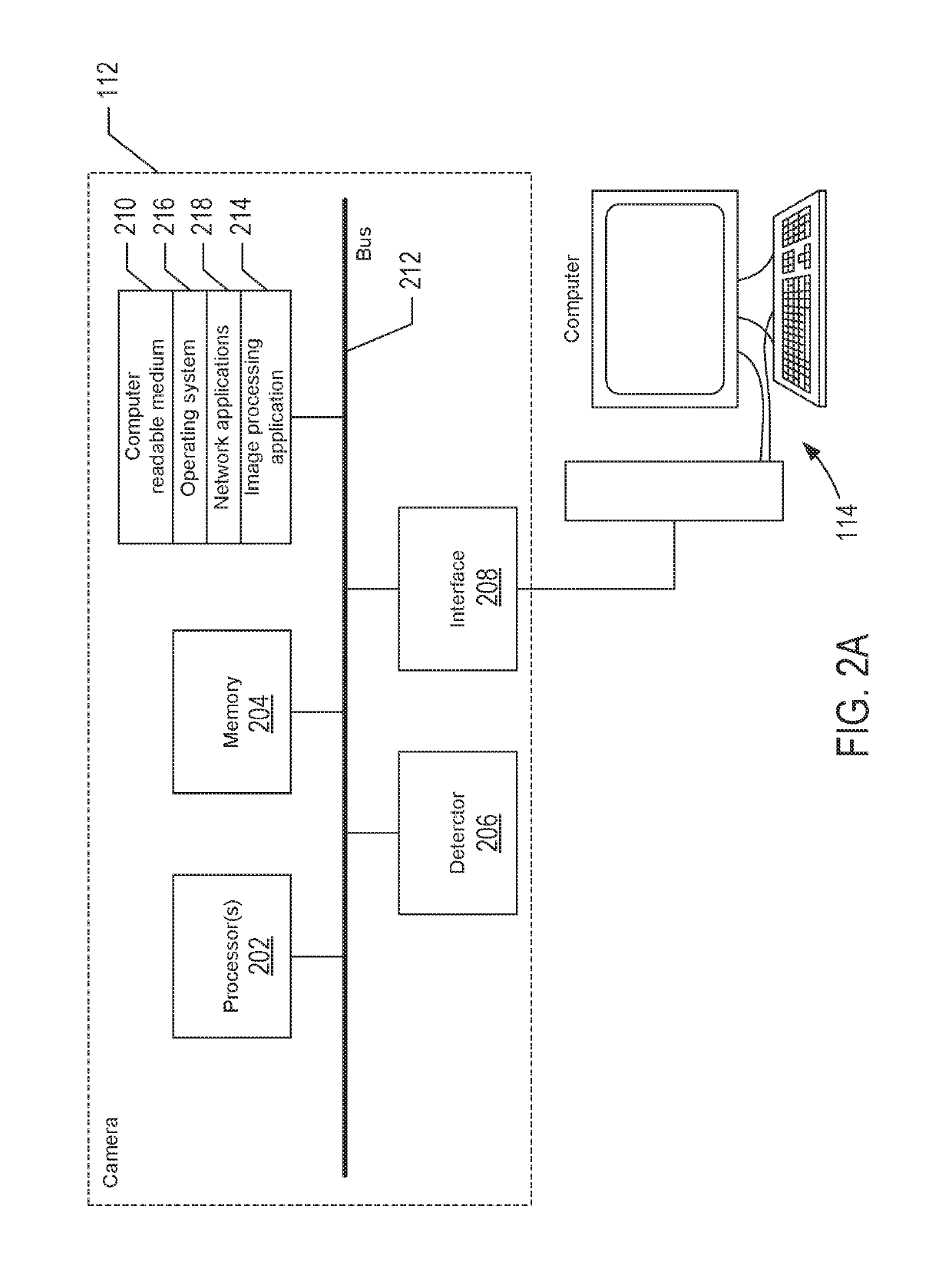
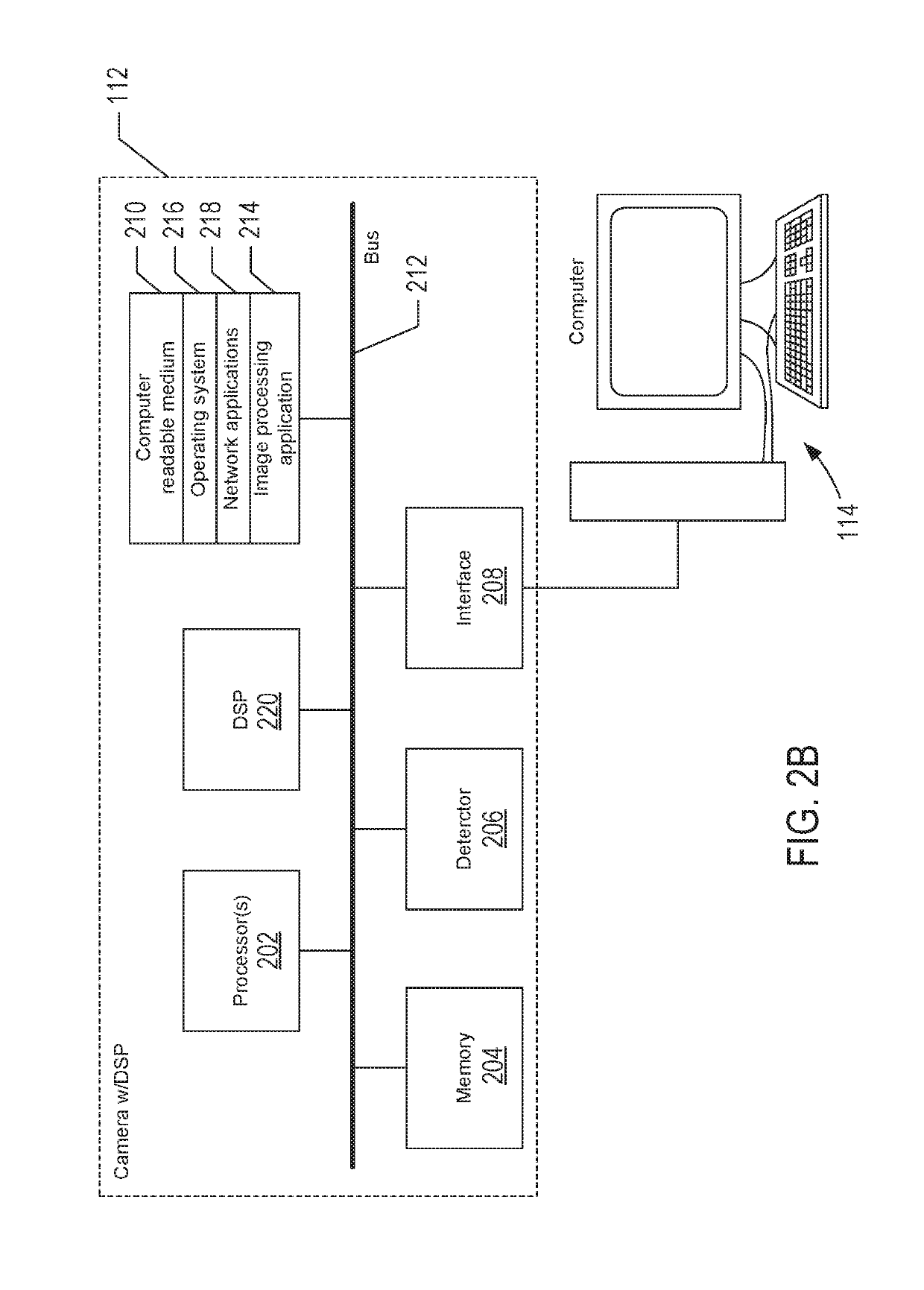
[0015]Systems and methods for executing super-resolution microscopy of specimen within the camera of a fluorescence microscopy instrument greatly reduces the amount of data that is typically transferred to the instrument computer, which reduces the amount of data storage used by the computer and eliminates the need for a high-speed interface between the camera and the computer. In addition, by performing most of the image processing within the camera and avoiding a large number of data transfers from the camera to the computer, the overall performance of the fluorescence microscope instrument is improved.

[0016]FIG. 1 shows a schematic representation of an example fluorescence microscopy instrument 100 used to perform super-resolution microscopy. The instrument 100 includes a light source 102, an excitation filter 104, a dichroic mirror 106, an objective lens 108, an emission filter 110, a camera 112, and a computer 114. The light source 102 can be configured with a number of separat...


PUM


Abstract

Systems and methods for executing super-resolution microscopy of a specimen with most of the image processing performed in a camera of a fluorescence microscopy instrument are described. In one aspect, the camera includes one or more processors to execute machine-readable instructions that control excitation light output from a multi-channel light source, control capture of intermediate images of the specimen, and perform image processing of the intermediate images to produce a final super-resolution image of the specimen.

Description

CROSS-REFERENCE TO A RELATED APPLICATION[0001]This application is a Continuation of U.S. application Ser. No. 13 / 978,800, filed on Jul. 9, 2013, which is a filing under 35 U.S.C. 371 of International Application Number PCT / SE2012 / 050011, filed on Jan. 11, 2012, which claims priority to U.S. Provisional Patent Application No. 61 / 432,015, filed on Jan. 12, 2011. The entire teachings of the above applications are incorporated herein by reference.TECHNICAL FIELD[0002]This disclosure relates to fluorescence microscopy, and in particular, to systems and methods used to perform fluorescence microscopy.BACKGROUND[0003]In recent years, a number of techniques have been developed to increase the resolution of light microscopy beyond the diffraction limit. Some of these techniques involve stochastically activating the fluorescence of a subset of molecules that are present in a field-of view, capturing an image of those molecules that are activated with a digital camera, transferring the image t...

Claims


Application Information

Patent Timeline
IPC IPC(8): G02B21/16G01B11/00G02B21/02G01N21/64G02B21/36G06T3/40G02B21/06G02B21/08
CPCG01N21/6458G01B11/002G01N21/6408G01N21/6428G02B21/02G02B21/16G02B21/367G06T3/4053G02B21/361G02B21/082G01N2201/06113G01N2201/12G02B21/06
Inventor GOODWIN, PAUL C.
Owner LEICA MICROSYSTEMS CMS GMBH
Why Eureka
Try Eureka