
Cine phase contrast simultaneous multi-slice and multi-slab imaging of blood flow and cerebrospinal fluid motion

a technology of cerebrospinal fluid and contrast agent, which is applied in the field of magnetic resonance imaging, can solve the problems of inability to use intravenous contrast agent, inability to accurately measure velocity and blood flow, so as to reduce the artifactual lack of blood flow, reduce the effect of distance and accuracy degradation

Active Publication Date: 2019-03-26
ADVANCED MRI TECH
View PDF3 Cites 0 Cited by
  • Summary
  • Abstract
  • Description
  • Claims
  • Application Information

AI Technical Summary

Benefits of technology

This approach significantly enhances imaging speed and quality, reduces cardiac cycle-dependent artifacts, and improves spatial resolution by allowing greater inflow of unsaturated spins into the imaging volume, providing comprehensive coverage of blood flow in thicker slabs without contrast agents, thus overcoming limitations of conventional single-slice or single-slab imaging.

Problems solved by technology

As a consequence of either the velocity being too low or the slab being too thick, the branched vessels in the downstream regions of the body in the distal imaging slab may show a lack of signal, which is artifact.
This can degrade the accuracy of measuring velocity and blood flow.
This is particularly valuable because intravenous contrast agent can be a safety risk and may not be able to be used in patients who have compromised renal systems and are even at risk of renal failure from the contrast agent.
With the new method of simultaneous imaging, there is greater inflow of unsaturated spins into the volume undergoing repeated excitation and this makes the use of contrast agent unnecessary.
Second, these spins entering a thinner slab travel a shorter distance to fill even the distal regions of the thinner slabs.

Method used


Image

  • Cine phase contrast simultaneous multi-slice and multi-slab imaging of blood flow and cerebrospinal fluid motion
  • Cine phase contrast simultaneous multi-slice and multi-slab imaging of blood flow and cerebrospinal fluid motion
  • Cine phase contrast simultaneous multi-slice and multi-slab imaging of blood flow and cerebrospinal fluid motion

Examples


Embodiment Construction

[0018]A detailed description of examples of preferred embodiments is provided below. While several embodiments are described, the new subject matter in this patent specification is not limited to any one embodiment or combination of embodiments described herein but encompasses numerous alternatives, modifications, and equivalents. While numerous specific details are set forth in the following description to provide a thorough understanding, some embodiments can be practiced without some or all these details. For clarity, certain technical material that is known in the related art has not been described in detail to avoid unnecessarily obscuring the new subject matter described herein. Individual features of one or several of the specific embodiments described herein can be used in combination with features or other described embodiments. Further, like reference numbers and designations in the various drawings indicate like elements.

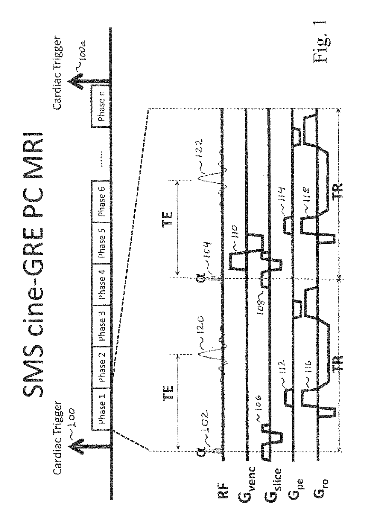
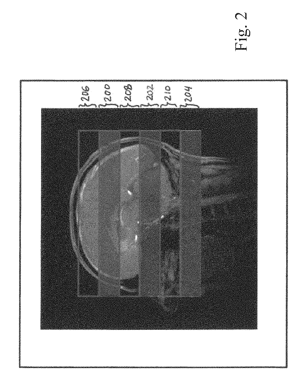
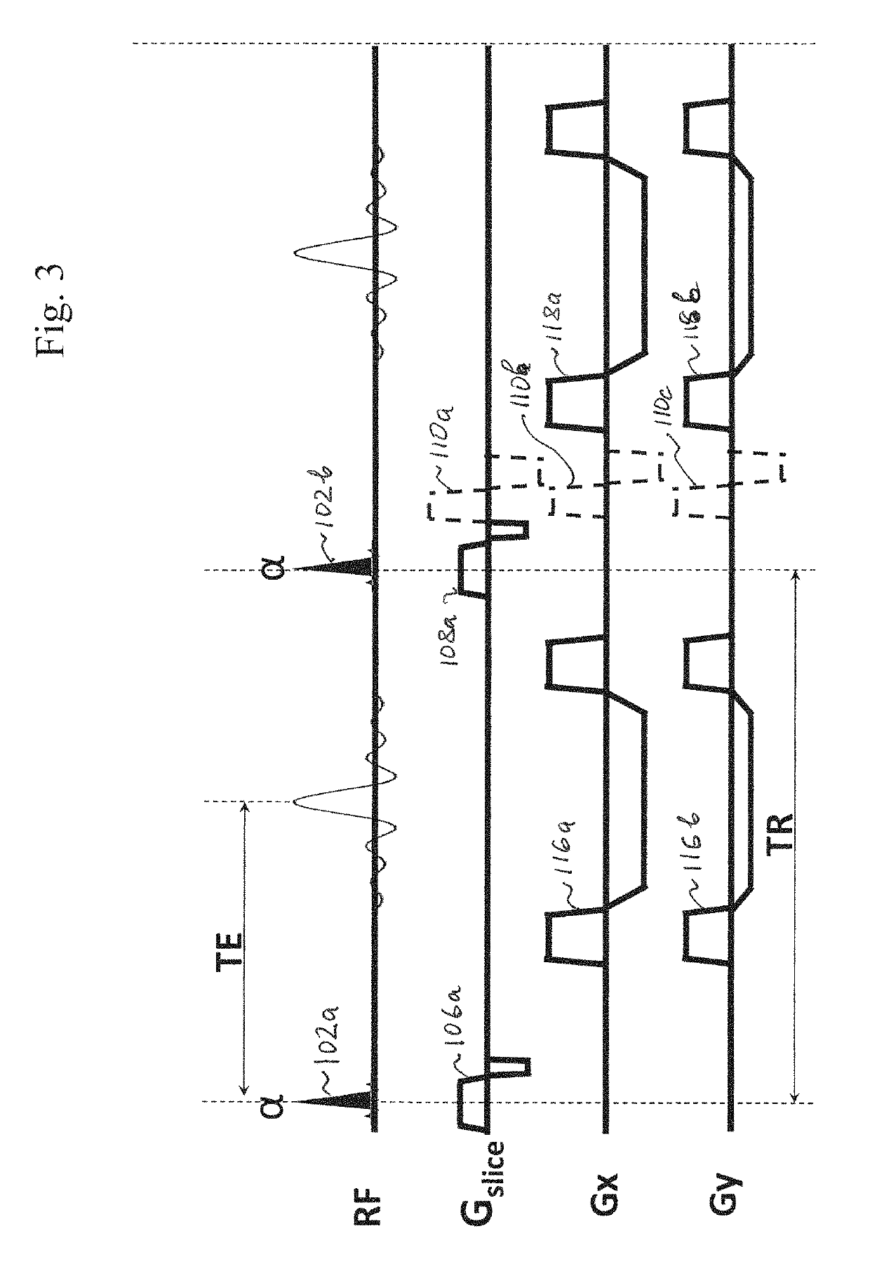
[0019]FIG. 1 illustrates a non-limiting example of ...


PUM


Abstract

An MRI method and an MRI system that simultaneously detect blood and / or CSF velocity or flow in plural slices or slabs preferably as not spatially adjacent. Two or more sets of interleaved slices or slabs can be assembled to cover the desired volume and derive higher quality MRI signals and images without a need for a contrast agent, even where the volume is too large for effective flow imaging with known techniques without contrast agent.

Description

REFERENCE TO RELATED APPLICATION[0001]This patent application claims the benefit of U.S. Provisional patent application 62 / 468,667 filed Mar. 8, 2017, and hereby incorporates by reference its entire contents.FIELD[0002]This patent specification relates to magnetic resonance imaging (MRI).BACKGROUND[0003]This patent specification refers to, and hereby incorporates by reference, the items listed at the end and identified by reference (ref) numbers in parenthesis in the discussion below.[0004]Phase contrast imaging is a magnetic resonance imaging (MRI) technique that can be used to visualize moving fluid. It is typically used for MR angiolgraphy that does not require introducing a contrast agent in the subject. Phase contrast imaging relies on the assumption that spins that are moving in the same direction as a magnetic field gradient develop a phase shift that that is proportional to the velocity of the spins. In principle, bipolar gradients (two gradients with equal magnitude but opp...

Claims


Application Information

Patent Timeline
Patent Type & Authority Patents(United States)
IPC IPC(8): G01R33/563G01R33/483G01R33/46A61B5/055A61B5/145A61B5/00
CPCG01R33/56325A61B5/055A61B5/14507G01R33/4633G01R33/4835G01R33/56316A61B5/4076G01R33/56308A61B5/7285A61B5/026
Inventor FEINBERG, DAVIDCHEN, LIYONG
Owner ADVANCED MRI TECH
Learn More