Methods for forming three-dimensional human retinal tissue in vitro

a three-dimensional human retinal tissue and stem cell technology, applied in the field of stem cells, can solve the problems of in vitro preparation, structural and molecular features characteristic of advanced photoreceptor differentiation, and have not yet been observed, and achieve the effect of high-simplification

Active Publication Date: 2019-10-08
THE JOHN HOPKINS UNIV SCHOOL OF MEDICINE
View PDF3 Cites 0 Cited by
  • Summary
  • Abstract
  • Description
  • Claims
  • Application Information

AI Technical Summary

Benefits of technology

[0005]Many forms of blindness result from the dysfunction or loss of retinal photoreceptors1. Stem cells, especially induced pluripotent stem cells (iPSCs), may hold great promise for the modeling and / or therapy of diseases2-4. Previous work has shown that, when provided with the appropriate cues, mouse and human embryonic stem (ES) cells in culture can develop into a 3-dimensional eyecup that remarkably resembles the vertebrate eye, including differentiation of photoreceptor-like cells5,6. Nonetheless, structural and molecular features characteristic of advanced photoreceptor differentiation, such as the formation of outer-segment discs and photosensitivity in these in vitro preparations, have not yet been observed. The present inventors report here that, with a new, highly-simplified procedure, hiPSCs in vitro can recapitulate spatiotemporally each of the main steps of retinal development in vivo and form 3-dimensional retinal cups. Moreover, the photoreceptors in our hiPSC-derived retinal tissue achieve advanced maturation, showing the beginning of outer-segment-disc formation and photosensitivity.

Problems solved by technology

Nonetheless, structural and molecular features characteristic of advanced photoreceptor differentiation, such as the formation of outer-segment discs and photosensitivity in these in vitro preparations, have not yet been observed.

Method used


Image

  • Methods for forming three-dimensional human retinal tissue in vitro
  • Methods for forming three-dimensional human retinal tissue in vitro
  • Methods for forming three-dimensional human retinal tissue in vitro

Examples


examples

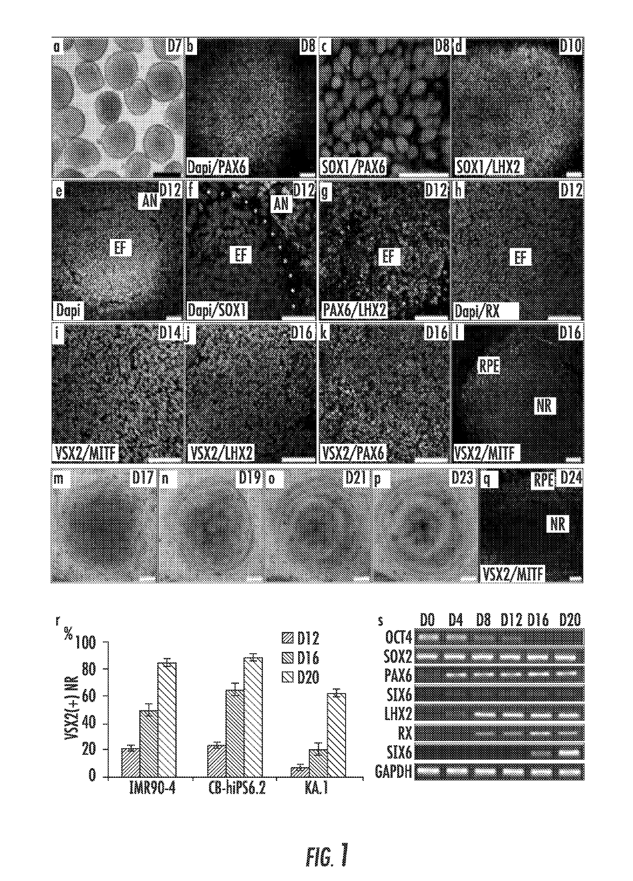
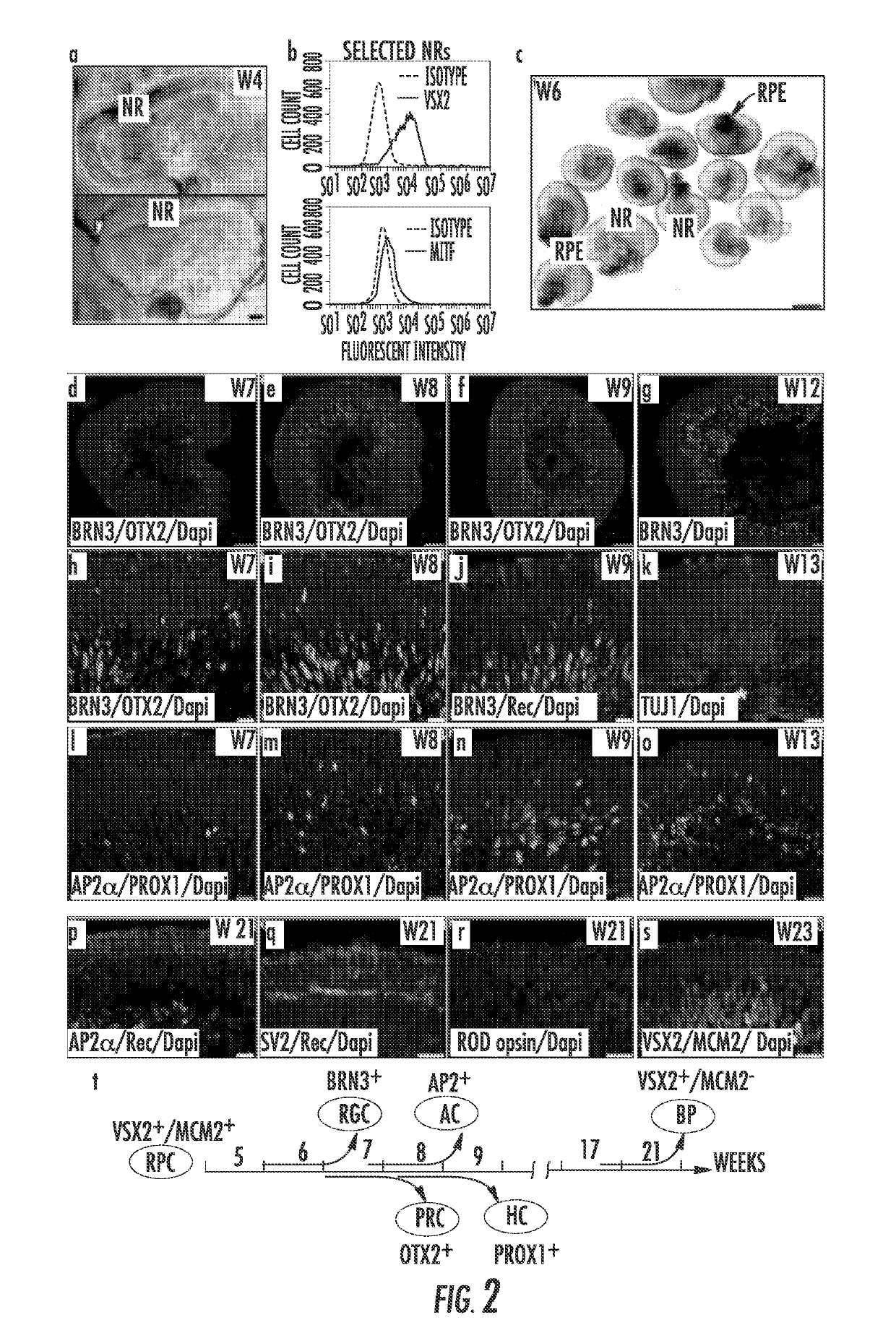
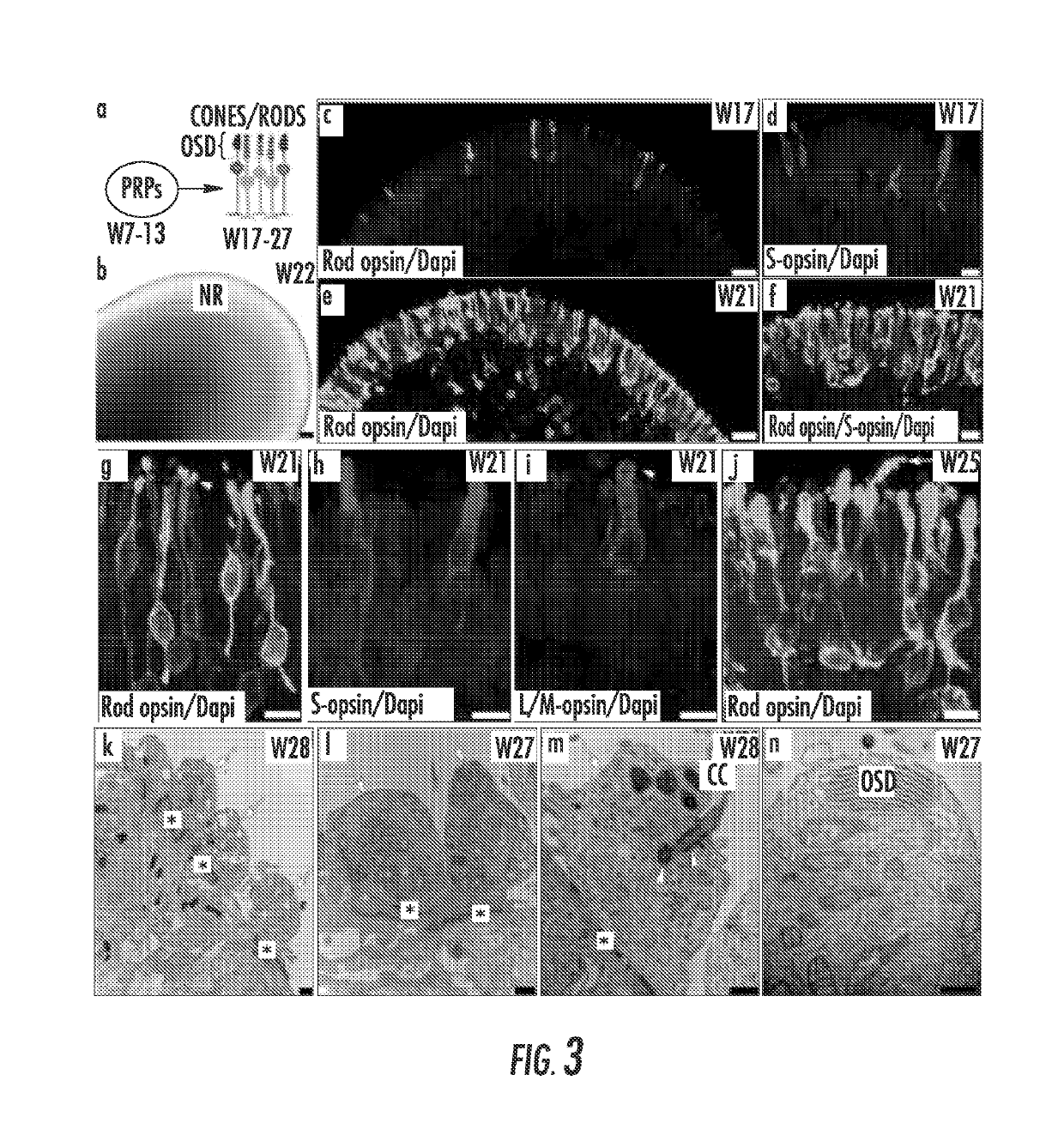
[0045]The following examples are put forth so as to provide those of ordinary skill in the art with a complete disclosure and description of how the compounds, compositions, articles, devices, and / or methods described and claimed herein are made and evaluated, and are intended to be purely illustrative and are not intended to limit the scope of what the inventors regard as their invention. Efforts have been made to ensure accuracy with respect to numbers (e.g., amounts, temperature, etc.) but some errors and deviations should be accounted for herein. Unless indicated otherwise, parts are parts by weight, temperature is in degrees Celsius or is at ambient temperature, and pressure is at or near atmospheric. There are numerous variations and combinations of reaction conditions, e.g., component concentrations, desired solvents, solvent mixtures, temperatures, pressures and other reaction ranges and conditions that can be used to optimize the product purity and yield obtained from the d...


PUM

PropertyMeasurementUnit
time-aaaaaaaaaa
time-aaaaaaaaaa
pHaaaaaaaaaa

Abstract

The present invention relates to the field of stem cells. More specifically, the invention provides methods and compositions useful for forming three-dimensional human retinal tissue in vitro. In a specific embodiment, an in vitro method for differentiating hiPSCs into three-dimensional retinal tissue comprising functional photoreceptors comprises the steps of (a) culturing the hiPSCs to form aggregates; (b) transitioning the aggregates into a neural induction medium; (c) seeding the aggregates on to extracellular matrix coated cell culture substrates; (d) replacing NIM with a chemically-defined differentiation medium; (e) detaching NR domains; (f) culturing in suspension; and (g) adding animal serum or plasma component and retinoic acid.

Description

CROSS-REFERENCE TO RELATED APPLICATIONS[0001]This application is a 35 U.S.C. § 371 U.S. national entry of International Application PCT / US2015 / 011701, having an international filing date of Jan. 16, 2015, which claims the benefit of U.S. Provisional Application No. 61 / 928,116, filed Jan. 16, 2014, the content of each of the aforementioned applications is herein incorporated by reference in their entirety.FIELD OF THE INVENTION[0002]The present invention relates to the field of stem cells. More specifically, the invention provides methods and compositions useful for forming three-dimensional human retinal tissue in vitro.INCORPORATION-BY-REFERENCE OF MATERIAL SUBMITTED ELECTRONICALLY[0003]This application contains a sequence listing. It has been submitted electronically via EFS-Web as an ASCII text file entitled “P12243-02.txt.” The sequence listing is 3,331 bytes in size, and was created on Jan. 16, 2015. It is hereby incorporated by reference in its entirety.BACKGROUND OF THE INVEN...

Claims


Application Information

Patent Timeline
Patent Type & Authority Patents(United States)
IPC IPC(8): C12N5/0793
CPCC12N5/062C12N2506/45C12N2533/90C12N2501/385C12N2509/00
Inventor CANTO-SOLER, MARIA VALERIAZHONG, XIUFENG
Owner THE JOHN HOPKINS UNIV SCHOOL OF MEDICINE
Why Eureka
Try Eureka