
Computer tomography X-ray imaging

a computer and x-ray imaging technology, applied in the field of computer tomography x-ray imaging, can solve problems such as noise, and achieve the effect of improving the reconstruction data

Active Publication Date: 2019-11-26
KONINKLJIJKE PHILIPS NV
View PDF11 Cites 0 Cited by
  • Summary
  • Abstract
  • Description
  • Claims
  • Application Information

AI Technical Summary

Benefits of technology

[0014]Thereby, slice normalization is performed, for example to compensate for tube flux fluctuations. Advantageously, the slice normalization processing for the different energy ranges results in less difference between measurements, and leads to improved results when reconstructing the image on behalf of the corrected or normalized projection data. It also improves to reduce artifacts caused e.g. by outer detector elements being affected by the patient or any other object inside the system. Due to providing equal normalization, the need to check if an object influence is detected, which might lead to the need to turn off slice normalization procedure, which would result in abrupt changes in prepped data, the procedure based on the present invention is less prone to error and the provided data is more consistent data and artefacts are reduced.
[0016]The term “normalization” relates to a process in image processing where the range of pixel intensity values is changed. For example, the range is changed such that the pixel intensity values are aligned with a predetermined standard pixel intensity value for a predetermined radiation attenuation. In another example, the range is changed to improve further image processing procedures, for example, contrast can be enhanced by the normalization.
[0034]According to an aspect, equal normalization is provided for two (or more) energy ranges of projected image data. One option to achieve a consistent processing of the two detector layers is to use the mean of the measured data of outer detector elements of upper and lower detector layer to determine the slice normalization-rescaling for both layers. Another option is to use only the measured data of one of the two layers to determine the slice normalization-rescaling for both layers. The measured data forming the basis of the normalization may vary due to different X-ray intensity output of the X-ray source. As the data is measured in areas outside the part of the radiation radiating through the patient, the measured radiation can be taken and put into relation to a standardized intensity value. The resulting normalization thus considers a possible change in intensity of the radiation from the X-ray tube, such as caused by flux intensity variations of an X-ray tube, and results in improved data for the reconstruction.

Problems solved by technology

However, it has been shown that this may be subject to noise.

Method used


Image

  • Computer tomography X-ray imaging
  • Computer tomography X-ray imaging

Examples


Embodiment Construction

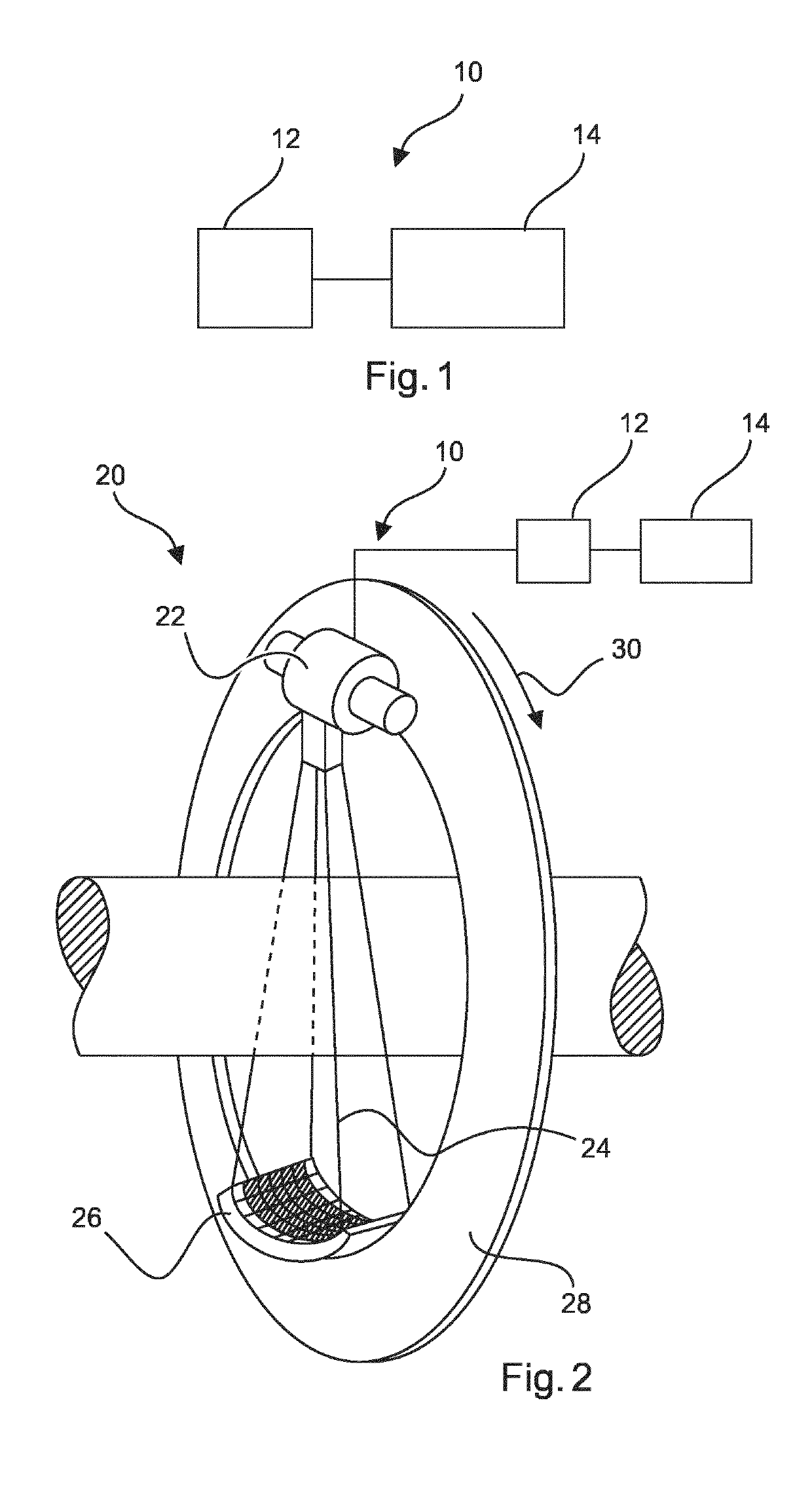
[0040]FIG. 1 shows a system 10 for computer tomography X-ray imaging. The system 10 comprises a data interface 12, and a processing unit 14. The data interface 12 is configured to provide at least first and second CT X-ray radiation projection data of an object for at least a first and second X-ray energy range. The ranges are different from each other. The at least first and second CT X-ray radiation projection data of the object comprises a plurality of slices of X-ray measurements from different angles to produce cross-sectional images of the object. The processing unit 14 is configured to determine a correction for slice normalization of the plurality of slices of X-ray measurements to change the first and the second CT X-ray projection data in terms of their pixel intensity values. The processing unit 14 is also configured to apply an equal slice normalization for the first and the second CT X-ray projection data, wherein the pixel intensity values of the plurality of slices of...


PUM


Abstract

The present invention relates to computer tomography X-ray imaging. In order to provide further improved data for the reconstruction, a system (10) for computer tomography X-ray imaging is provided. The system comprises a data interface (12) and a processing unit (14). The data interface is configured to provide at least first and second CT X-ray radiation projection data for at least a first and second X-ray energy range, which ranges are different from each other. The processing unit is configured to determine a correction for slice normalization, and to apply an equal slice normalization for the first and the second CT X-ray projection data and thereby generating prepared first and second CT X-ray projection data. For the correction, the equal slice normalization is based on measured data of outer detector elements. Further, the data interface is configured to provide the prepared first and second CT X-ray projection data for further processing. In an example, the system further comprises a computer tomography X-ray imaging acquisition arrangement (20) with an X-ray source (22) configured to generate an X-ray beam, and an X-ray detector (26) configured as an energy discriminating X-ray detector to simultaneously provide X-ray radiation projection data for at least two different X-ray energy ranges separately. The computer tomography X-ray imaging acquisition arrangement is configured to acquire at least the first and second CT X-ray projection data of a region of interest of an object for the at least first and second X-ray energy range.

Description

CROSS REFERENCE TO RELATED APPLICATIONS[0001]This application is the U.S. National Phase application under 35 U.S.C. § 371 of International Application No. PCT / EP2017 / 072039, filed Sep. 4, 2017, published as WO 2018 / 046414 on Mar. 15, 2018 which claims the benefit of European Patent Application Number 16187966.3 filed Sep. 9, 2016. These applications are hereby incorporated by reference herein.FIELD OF THE INVENTION[0002]The present invention relates to a system for computer tomography X-ray imaging and a method for computer tomography X-ray imaging as well as to a computer program element for controlling an apparatus and a computer readable medium.BACKGROUND OF THE INVENTION[0003]In spectral computer tomography (CT), X-ray radiation is detected for several energy levels or ranges to provide enhanced image projection data for reconstructing an image. In spectral CT imaging, when combining several projection data images, as a pre-processing procedure, an adaptation may be provided. F...

Claims


Application Information

Patent Timeline
Patent Type & Authority Patents(United States)
IPC IPC(8): G06K9/40A61B6/00G06T11/00G06K9/00A61B6/03
CPCA61B6/5211A61B6/032A61B6/4241G06T11/003A61B6/482G06T2211/408G06T2207/10081A61B6/5258
Inventor BRENDEL, BERNHARD JOHANNESKOEHLER, THOMASPROKSA, ROLAND
Owner KONINKLJIJKE PHILIPS NV
Learn More