

Performing segmentation of cells and nuclei in multi-channel images
a multi-channel image and cell technology, applied in image analysis, image enhancement, instruments, etc., can solve the problems of distorted image features being used for further image analysis or classification, unnatural transition between foreground and background, identification of artificial objects that may be difficult to discriminate, etc., to minimize side effects
- Summary
- Abstract
- Description
- Claims
- Application Information
AI Technical Summary
Benefits of technology
Problems solved by technology
Method used
Image

Examples
example 1
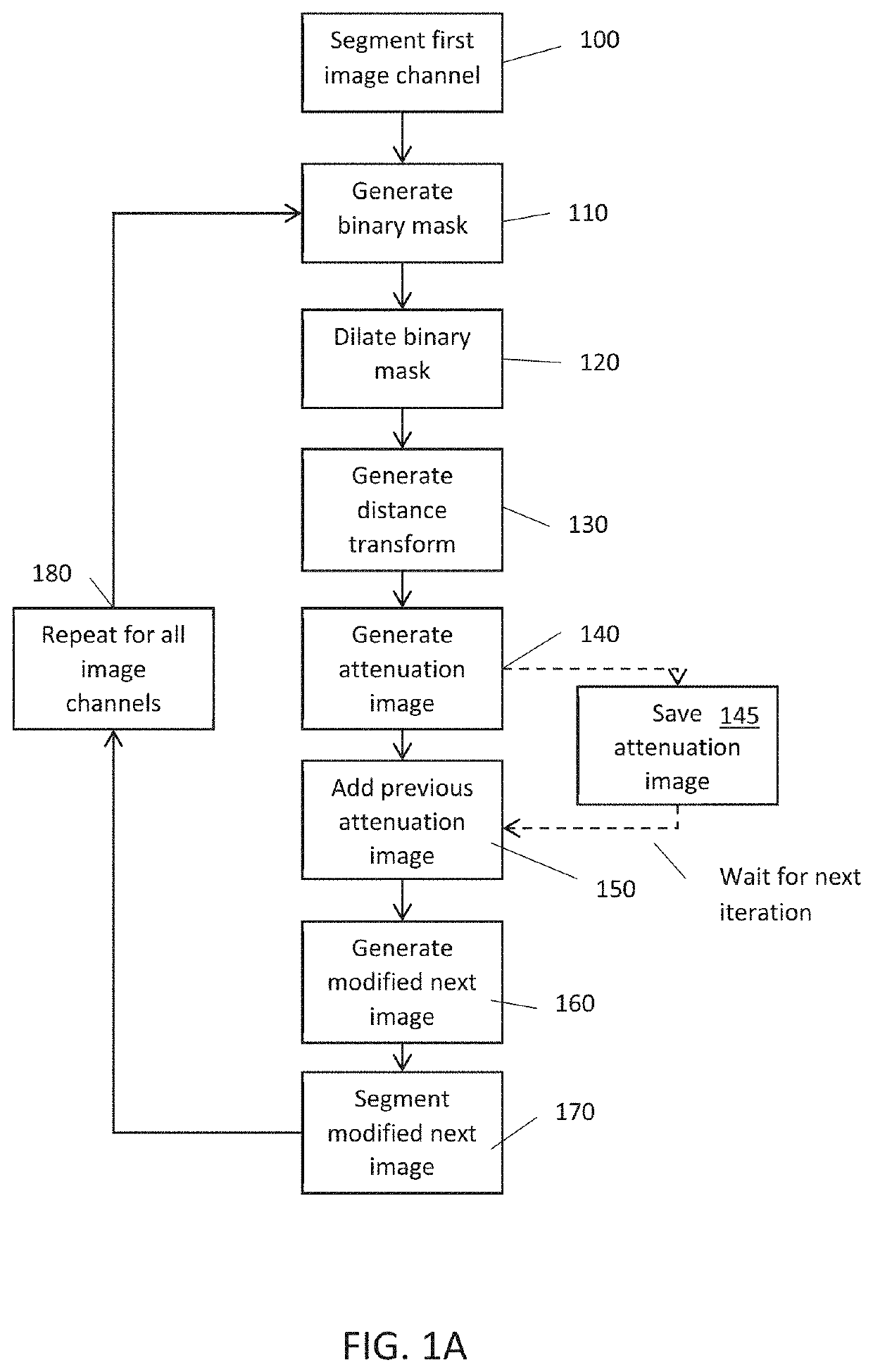
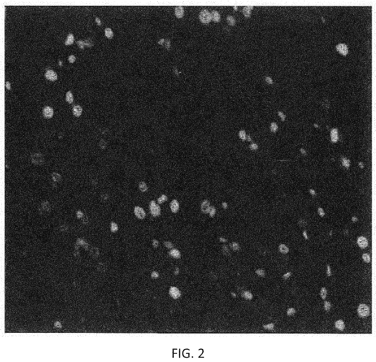
[0080]The input image set comprises a two-channel image. The two channels correspond to I1 and I2 as shown in FIG. 2 and FIG. 3. Image I1 (201) is segmented (100) and a binary image mask (202) is generated (110) and subsequently dilated (120) to cover the possible edge artifacts of the segmented regions. The binary image mask of nuclei segmented from I1, is shown in FIG. 4.
[0081]The dilated binary image mask (202) is then used to generate a distance transform image (203), by computing the distance to the edge of the masked (white) region for each pixel in the image (distance is zero outside the masked region)(130). The distance transform of the binary image mask is shown in FIG. 5.
[0082]The distance transform image (203) is then used to generate an attenuation image (204) by performing a polynomial transform (140) on each pixel of the distance transform image. The attenuation image is shown in FIG. 6. The polynomial transform in this example is a second-order polynomial parabola (m=...
PUM
 Abstract
Description
Claims
Application Information