Culture medium for expanding breast epithelial stem cells

a technology culture media, which is applied in the field of breast epithelial stem cell culture media and methods, can solve the problems of inability to support the long-term culture of epithelial stem cells or tissue fragments derived from breast tissue, inability to obtain large numbers of stem cells using this technique, and inability to support long-term culture of breast organoids

Active Publication Date: 2021-03-30
KONINK NEDERLANDSE AKADE VAN WETENSCHAPPEN
View PDF76 Cites 1 Cited by
  • Summary
  • Abstract
  • Description
  • Claims
  • Application Information

AI Technical Summary

Benefits of technology

[0197]Cells taken directly from live tissue, i.e. freshly isolated cells, are also referred to as primary cells. In some embodiments the epithelial stem cells are primary epithelial stem cells. Primary cells represent the best experimental models for in vivo situations. In a preferred embodiment of the invention, the epithelial stem cells are (or are derived from) primary epithelial stem cells. Primary cell cultures can be passaged to form secondary cell cultures. With the exception of cancer cells, traditional secondary cell cultures have a limited lifespan. After a certain number of population doublings (e.g. 50-100 generations) cells undergo the process of senescence and stop dividing. Cells from secondary cultures can become immortalized to become continuous cell lines. Immortalization can occur spontaneously, or may be virally- or chemically-induced. Immortalized cell lines are also known as transformed cells. By contrast, the methods of the present invention allow continuous passaging of epithelial stem cells without immortalisation or transformation. Thus in some embodiments, the epithelial stem cells are not immortalised or transformed cells or are not derived from an immortalised cell line or a transformed cell line. An advantage of the present invention is that the epithelial stem cells, undergoing multiple rounds of expansion and passaging, retain the characteristics of primary cells and have minimal or no genotypic or phenotypic changes.
[0198]The epithelial stem cells may be obtained by any suitable method. In some embodiments, cells are isolated by collagenase digestion, for example, as described in the examples and in Dorell et al., 2008 (Hepatology. 2008 October; 48(4):1282-91. Surface markers for the murine oval cell response. Dorrell C, Erker L, Lanxon-Cookson K M, Abraham S L, Victoroff T, Ro S, Canaday P S, Streeter P R, Grompe M). In some embodiments, collagenase digestion is performed on a tissue biopsy. In some embodiments, collagenase and accutase digestion are used to obtain the epithelial stem cells for use in the invention.
[0199]In some embodiments, breast tissue may be minced, washed (for example in basal culture medium) and incubated in a culture medium of the invention supplemented with a protease (for example, collagenase, dispase or trypsin) (see, for example,

Problems solved by technology

However, these methods have been found to be unable to support the long-term culture of epithelial stem cells or tissue fragments derived from breast tissue.
However, this system is unable to support long-term culture of breast organoids.
Accordingly, it is not possible to obtain large numbers of stem cells using this technique.
However, the authors of this paper provided no guidance with respect to culturing breast epithelial stem cells obtained from normal breast tissue, primary tumour or metastases.

Method used


Image

  • Culture medium for expanding breast epithelial stem cells
  • Culture medium for expanding breast epithelial stem cells
  • Culture medium for expanding breast epithelial stem cells

Examples


example 1

ocessing and Mammary Organoid Culture

Overview

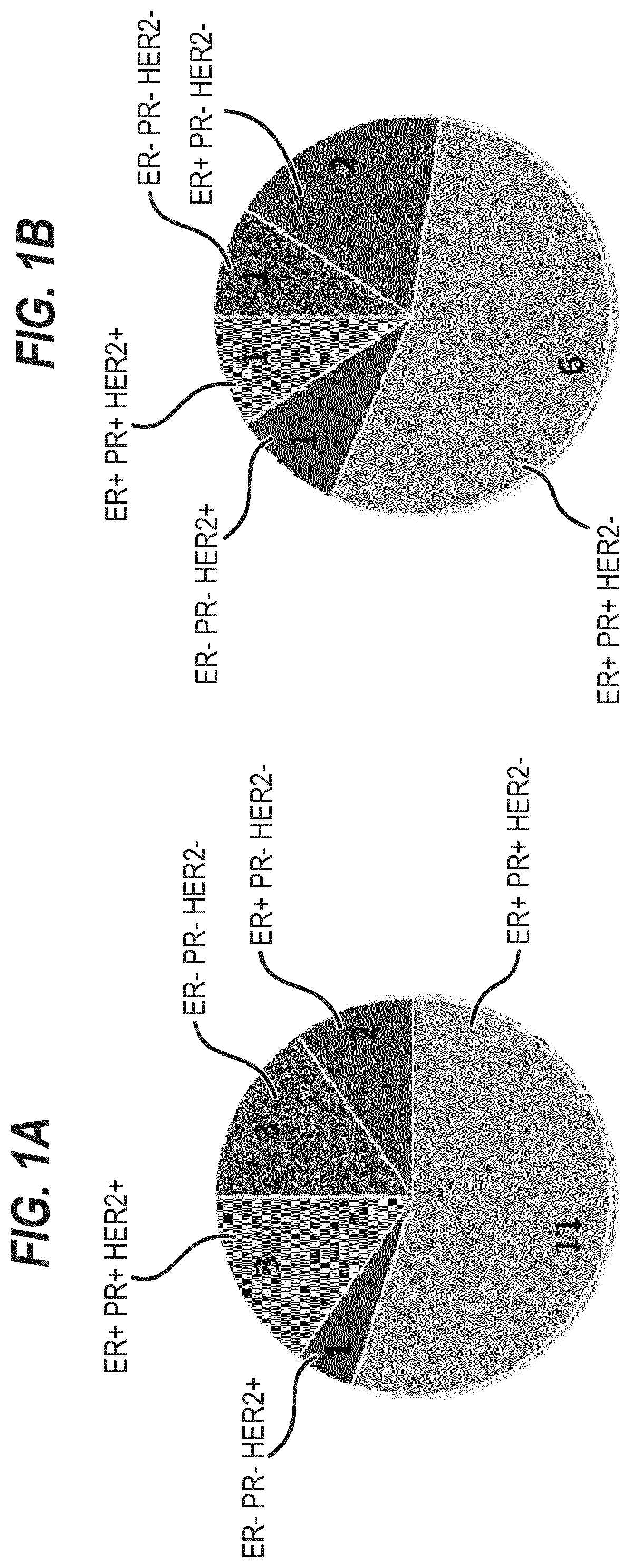
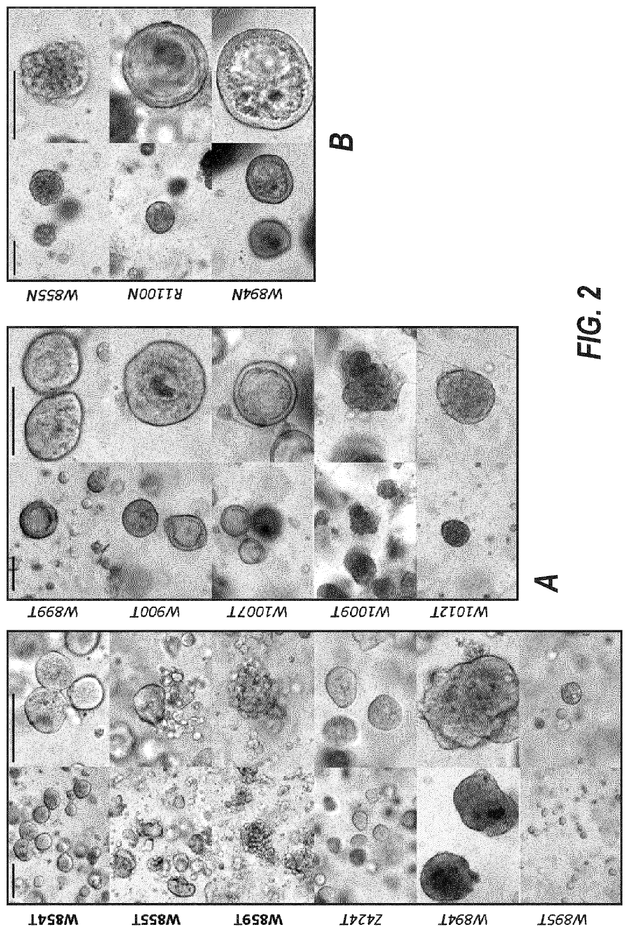
[0367]20 breast tumors were sampled, three of which were triple negative (15%, FIG. 1A and FIG. 13). We successfully established three tumor organoid lines (splitting ratio >1:2, passage >7). 8 additional cultures are currently expanding and will, based on our experience, most likely generate successful organoid lines (FIG. 2 and FIG. 13). A selection for a specific subtype is not evident (FIG. 1B).

[0368]From the 19 normal samples obtained we thus far established three promising cultures (FIG. 2, FIG. 13).

Culture Conditions

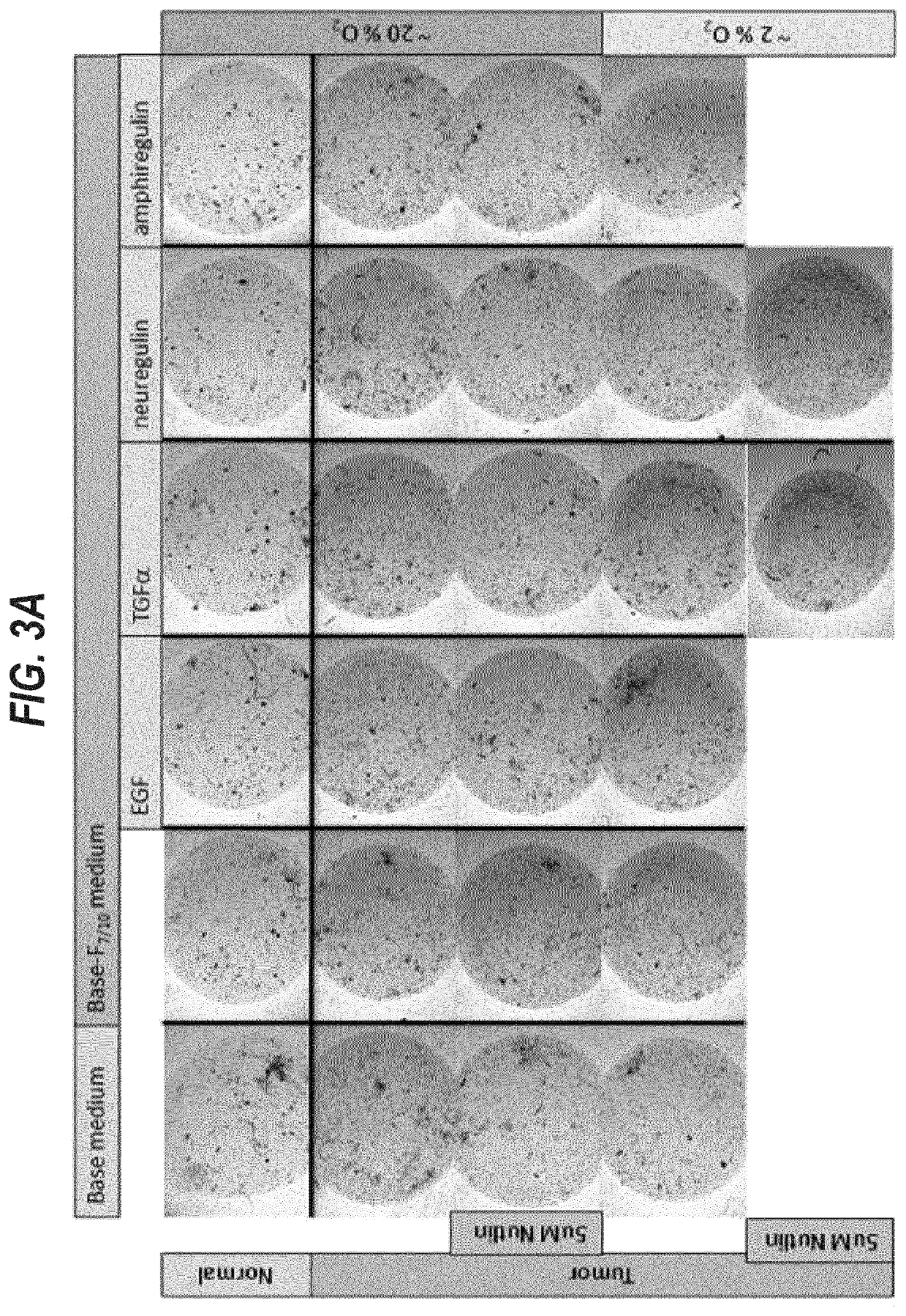
[0369]The first 12 samples were used to optimize cell isolation and organoid culture conditions. Samples 13-20 were grown in six different conditions.

Altered Parameters for Cell Isolation

[0370]For the isolation of viable cells we tested several digestive enzymes (collagenase, dispase, trypsin) at different concentrations (1-5 mg / ml) in different media (AdDF+++, base medium, base-F7 / 10N medium) for different periods of t...

example 2

ization of Established Mammary Tumor Organoid Lines

[0374]Mammary tumor organoid lines W854T, W855T, and W859T readily expand in base-F7 / 10N medium and were therefore characterized in more detail.

[0375]Their tumor status was confirmed through karyotyping revealing aneuploidy at varying degrees (FIG. 4).

[0376]Line W854T furthermore readily grew in medium containing 5 μM Nutlin-3 indicating the presence of mutant p53. RNA and DNA were isolated and sent to BI for gene expression profiling and exome sequencing.

[0377]Histological and immunofluorescent analysis confirmed that ER, PR, and HER2 status are conserved between originating tumor and established tumor organoid line after >7 passages (FIGS. 5 and 6).

[0378]Whereas normal organoid line W855N consists of mutually exclusive basal and luminal cells, tumor organoids solely express luminal cell marker Keratin-8 (FIGS. 6 and 7). E-cadherin expression is present but decreased in tumor compared to normal organoid lines indicating their epith...

example 3

ghput Drug Screen

[0379]To test the feasibility of mammary tumor organoids for drug screening, lines W854T, W855T, and W859T were plated in duplicate into a 96-well format and exposed to medium containing the EGFR inhibitor Iressa (1 nM-30 μM at 1:3 dilution steps, S1025 Selleck Chemicals) or the p53 stabilizer Nutlin-3 (1 nM-30 μM at 1:3 dilution steps, 10004372 Cayman Chemicals) for 7 days. Organoids were photographed every 3-4 days (FIG. 8) and cell viability measured at day 7 using the CellTiter-Glo 2.0 assay (G9241, Promega) (FIG. 9).

[0380]All three organoid lines respond to Iressa at concentrations ˜10 μM indicating a dependence on EGFR signaling. With perhaps the exception of W855T, Nutlin-3 does not inhibit growth at concentrations up 30 μM indicating the presence of mutant p53 (FIG. 9). Mammary tumor organoids are suitable for cell viability based low throughput drug screens in a 96-well format. We are currently testing the assay in a 384-well plate format which has been suc...


PUM

PropertyMeasurementUnit
widthsaaaaaaaaaa
widthsaaaaaaaaaa
concentrationaaaaaaaaaa

Abstract

The invention relates to improved culture methods for expanding epithelial stem cells and obtaining organoids, to culture media involved in said methods, and to uses of said organoids.

Description

[0001]This application is a national phase entry pursuant to 35 U.S.C. § 371 of International Application No. PCT / EP2015 / 077988, filed Nov. 27, 2015, which claims the benefit of priority of Great Britain Application No. 1421094.2, filed Nov. 27, 2014, each of which is incorporated by reference herein in its entirety for any purpose.[0002]All documents cited herein are incorporated by reference in their entirety.TECHNICAL FIELD[0003]The invention is in the field of breast epithelial stem cell culture media and methods, in particular culture media and methods for expanding populations of breast epithelial stem cells, e.g. human breast epithelial stem cells.BACKGROUND[0004]There is great interest in culture media and methods for expanding populations of stem cells. Populations of stem cells have many uses. For example, stem cells and their differentiated progeny can be used in cellular assays, drug screening, and toxicity assays. Stem cells also show promise for cell-based therapies, s...

Claims


Application Information

Patent Timeline
Patent Type & Authority Patents(United States)
IPC IPC(8): C12N5/00C12N5/095C12N5/071
CPCC12N5/0695C12N5/0631C12N5/00C12N2501/115C12N2501/415
Inventor SACHS, LARS NORMANGRACANIN, ANADROST, JARNO
Owner KONINK NEDERLANDSE AKADE VAN WETENSCHAPPEN
Why Eureka
Try Eureka