
Airway management device for identification of tracheal and/or esophageal tissue

a management device and esophageal technology, applied in the field of respiratory medical devices, can solve the problems of failure to intubate, death through hypoxemia or through unrecognized intubation of the esophagus, and still encounter major adverse events

Active Publication Date: 2021-04-20
UNITED STATES OF AMERICA THE AS REPRESENTED BY THE SEC OF THE ARMY
View PDF19 Cites 0 Cited by
  • Summary
  • Abstract
  • Description
  • Claims
  • Application Information

AI Technical Summary

Benefits of technology

The device enables accurate and continuous confirmation of tracheal tube placement, reducing the risk of misplacement and adverse events by utilizing reflectance spectra analysis, even in difficult airway conditions.

Problems solved by technology

Failed intubations can lead to death through hypoxemia or through unrecognized intubation of the esophagus.
Even successful intubations, if delayed or performed poorly, can still encounter major adverse events, such as cardiac arrest, hypotension, hypoxemia or asphyxiation due to patient aspiration.
In addition to the number of attempts, the time to intubation also adds risk to patient outcomes with longer intubation times leading to extended oxygen deprivation, which has been associated with worse long-term outcomes for traumatic brain injury patients.
While visualization of the endotracheal tube passing through the vocal cords is considered the “gold standard” of intubation confirmation, visual depth into the patient's upper airway can be severely limited due to their clinical condition or innate anatomical characteristics.
These challenges, such as large or swollen tongue, short neck, prominent teeth, or maxillofacial trauma, are categorized as a “difficult airway” and pose problems for standard intubation and visual confirmation techniques.
However, these methods may require the provider to stop care to perform the confirmation or they may require additional specialized equipment that is not always readily available.
Furthermore, not all of the current methods, such as the EDD or visualization, provide continuous patient monitoring to ensure proper tracheal tube placement once intubated.
Displacement of the tracheal tube can result from patient movement and can easily be overlooked or go undetected amidst the typically chaotic prehospital, ICU and emergency departments and lead to the same serious adverse events.

Method used


Image

  • Airway management device for identification of tracheal and/or esophageal tissue
  • Airway management device for identification of tracheal and/or esophageal tissue
  • Airway management device for identification of tracheal and/or esophageal tissue

Examples


Embodiment Construction

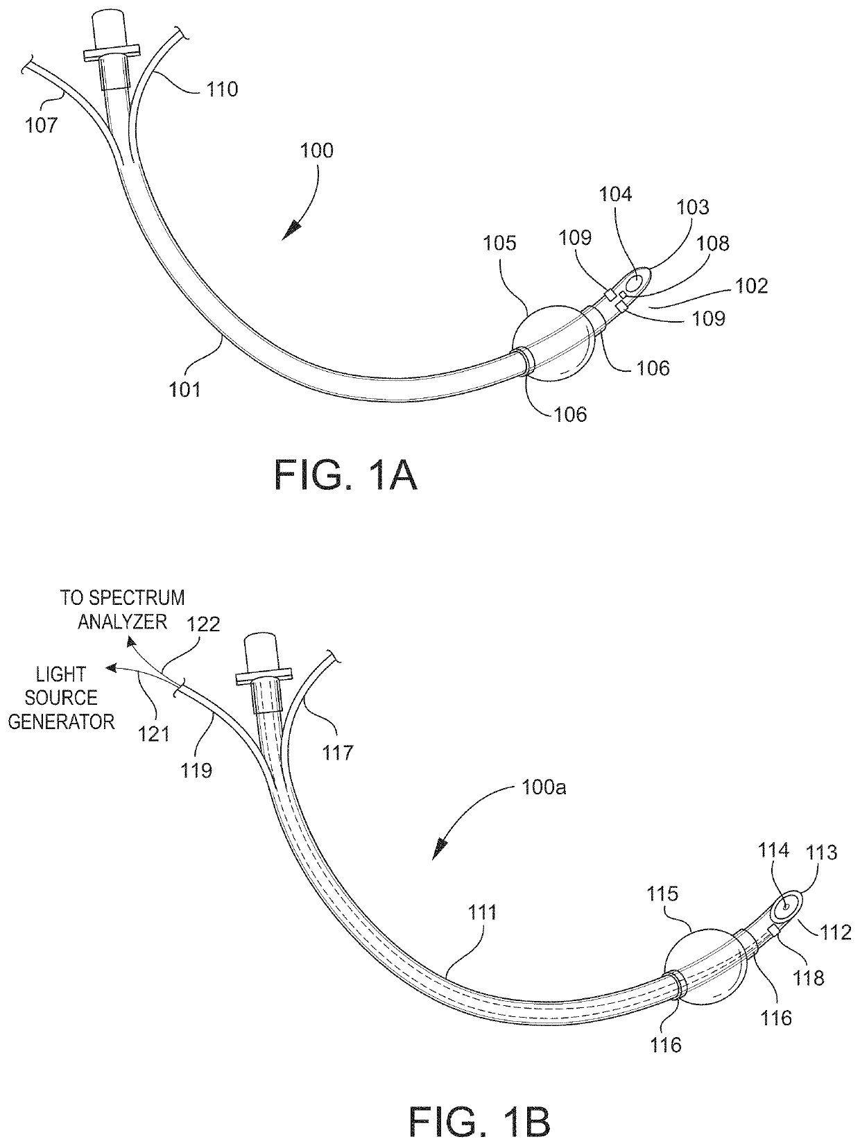
[0056]A novel airway management device may be used on subjects such as humans and animals. The device includes an airway tube with a first end that is disposed external to the subject and a second end that is disposed in a portion of the subject's airway. The device may take many forms, for example, the device may be in the form of a bougie, tracheal tube, tracheostomy tube, laryngeal mask, or supraglottic airway devices.

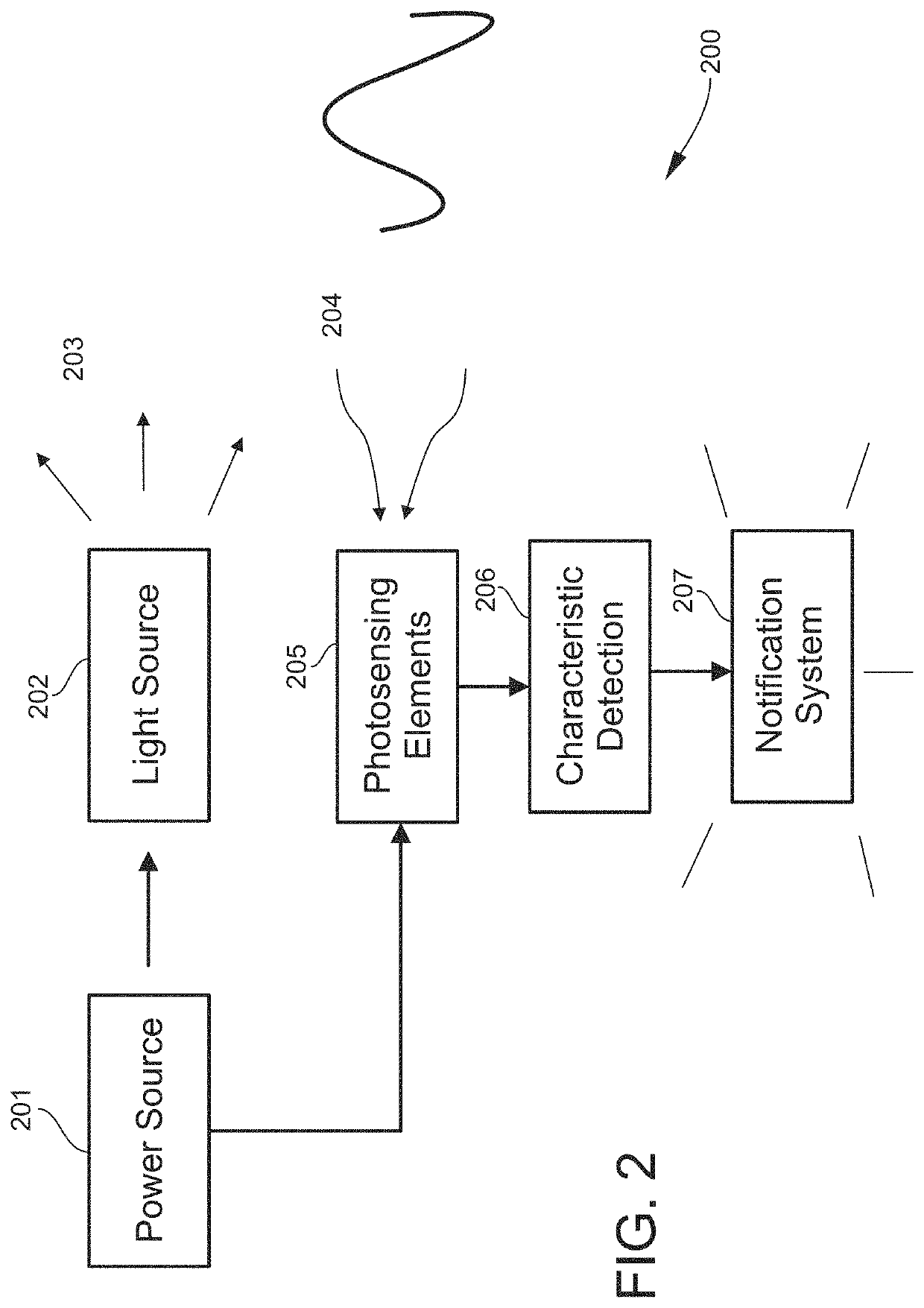
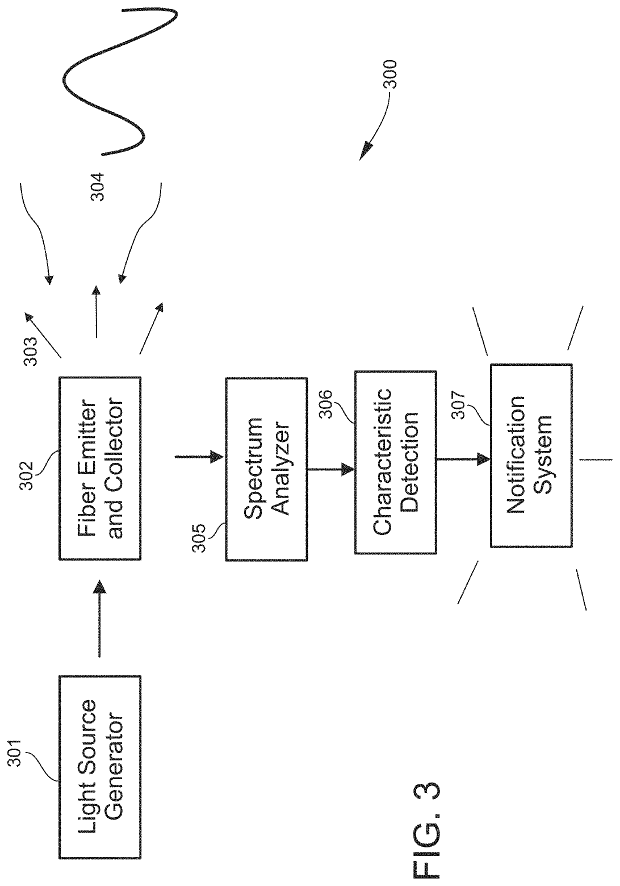
[0057]In one embodiment, the device may be used to positively identify tracheal tissue in the subject. In another embodiment, the device may be used to positively identify esophageal tissue in the subject. In a further embodiment, the device may be used to identify one or both of tracheal tissue and esophageal tissue in the subject. The identification of tracheal and / or esophageal tissue may be based on the optical reflectance spectrum of the particular tissue. In one embodiment, the optical reflectance spectrum is in the visible light range.

[0058]FIG. 1A shows one ...


PUM


Abstract

An airway management device (10) for a human or animal subject (20) includes an airway tube (101) having a first end for disposal external to the subject (20) and a second end (102) for disposal in a portion of an airway of the subject (20). A light emitting element (108) and a photo-sensing element (109) may be disposed at the second end (102) of the airway tube (101). The light emitting element (108) may be configured to transmit light to tissue adjacent to the light emitting element (108). The photo-sensing element (109) may be configured to receive reflectance spectra (204) from the tissue. The location of the second end (102) of the airway tube (101) may be determined from characteristics of the reflectance spectra (204) of the tissue.

Description

CROSS REFERENCE TO RELATED APPLICATIONS[0001]The present application claims the benefit of priority of Patent Cooperation Treaty international patent application number PCT Application No. PCT / US17 / 16174 filed on Feb. 2, 2017 and U.S. provisional patent application Ser. No. 62 / 292,277 filed on Feb. 6, 2016, both of which are expressly incorporated by reference herein.STATEMENT OF GOVERNMENT INTEREST[0002]The invention described herein may be manufactured, used and licensed by or for the United States Government.BACKGROUND OF THE INVENTION[0003]The invention relates in general to respiratory medical devices and in particular to airway management devices.[0004]Endotracheal intubation (ETI) is a frequent life-saving medical procedure. ETI is often performed in a prehospital or emergency medicine environment when a patient cannot sufficiently breathe of their own accord. Common medical emergencies such as cardiac arrest or trauma often necessitate paramedics or nurses to perform ETI in ...

Claims


Application Information

Patent Timeline
Patent Type & Authority Patents(United States)
IPC IPC(8): A61M16/04A61B1/267A61B5/06A61B5/00A61B5/1459A61B5/08A61B5/083
CPCA61M16/045A61B1/267A61B5/0075A61B5/0084A61B5/065A61B5/1459A61M16/0411A61M16/0488A61B5/08A61B5/0836A61B5/4887A61M16/0418A61M16/0434A61M2205/3313A61M2205/581A61M2205/583
Inventor NAWN, CORINNESOUHAN, BRIAN
Owner UNITED STATES OF AMERICA THE AS REPRESENTED BY THE SEC OF THE ARMY
Learn More