Screening method and apparatus for detecting an object of interest

a technology of object detection and screening method, applied in the direction of material analysis using wave/particle radiation, instruments, image enhancement, etc., can solve the problems of difficult recognition of fibers, non-uniform surface structure of the pores of the filter, etc., to improve the detection efficiency, easy and efficient detection, and the effect of detecting the manipulated imag

Active Publication Date: 2021-09-07
PHENOM WORLD HLDG BV
View PDF3 Cites 0 Cited by
  • Summary
  • Abstract
  • Description
  • Claims
  • Application Information

AI Technical Summary

Benefits of technology

[0008]According to the method, an image of said sample is acquired. In practice, this means that an image is obtained of at least part(s) of the sample carrier with its surface structure. It is recognized by the inventors that the surface structure of the sample carrier is visible in the acquired image, and that this hinders the detection of the object of interest. For example, the above mentioned asbestos detection method uses gold-coated porous filters. The pores of these filters provide a non-uniform surface structure. The pores are visible in the acquired image, and may obscure parts of the fibers. In addition, the pores provide an image that is relatively complex for quick interpretation, creating a vigilant task for the operator that makes recognition of fibers difficult. To this end, in the method of the disclosure it is proposed to provide information on said surface structure of said sample carrier, and to use this information for manipulating the acquired image. Manipulating means in particular reducing or removing the surface structure of the sample carrier, for creating an image of the sample wherein the surface structure is removed or less pronounced. Then, the manipulated image may screened for detecting said object of interest. Screening the manipulated image is easier and less strenuous, as the complexity of the image is reduced by at least partly removing the surface structure of the sample carrier. With this, the object as defined above is achieved.
[0010]The step of providing information on said surface structure may, in an embodiment, comprise the step of acquiring an image of said sample carrier. This may in particular include acquiring an image of the sample carrier alone, i.e. without (or substantially without) the object of interest. For example, it is conceivable that an image of a sample carrier is taken before (potentially) applying the object of interest thereto. Then, an image of the sample (i.e. comprising said same sample carrier but now—potentially—including the object of interest) is acquired as well. As the same carrier is imaged before and after, image manipulation may be used to (partly) remove the surface structure of the carrier from the image of the sample. This way, screening for the object of interest becomes easier.
[0024]Advantages of such an apparatus have been described above with respect to the method as disclosed herein. In particular, the apparatus allows easier and more efficient detection of objects of interests, by manipulating an acquired image, using information on the surface structure of the sample carrier. Said information on the surface structure may be provided beforehand, for example by providing an image of the surface structure, or by providing a model of the surface structure. Said image may be a pre-recorded image. Said model may contain mathematical and / or statistical information, for example.
[0025]It is envisaged that the apparatus as described herein allows human operators to screen the manipulated image more easily and effectively, as the manipulated image is visually less complex and therefore easier to interpret by a human operator. In an embodiment, however, said processing device is arranged for screening said manipulated image for said object of interest. This allows the manipulated image to be screened by means of image analysis techniques, such as, for example a neural network.
[0027]In a special embodiment, the apparatus is, or at least comprises, a charged particle microscope. In particular, the charged particle microscope may be a scanning electron microscope (SEM). The charged particle microscope may comprise a backscatter electron detector (BSD) that is arranged for acquiring said image of said sample, and may comprise a secondary electron detector (SED) that is arranged for acquiring said second image of said sample carrier. The BSD is in particular sensitive to the sample carrier including the object of interest, and the SED is in particular sensitive to the sample carrier and insensitive to the object of interest. Therefore, the image obtained with the SED can be used as information regarding the surface structure of the sample carrier for manipulating the image obtained with the BSD. One further advantage is that the image obtained with the SED and the image obtained with the BSD can be obtained substantially simultaneously, using a single scan—giving a high correlation between the two acquired images.

Problems solved by technology

The pores of these filters provide a non-uniform surface structure.
In addition, the pores provide an image that is relatively complex for quick interpretation, creating a vigilant task for the operator that makes recognition of fibers difficult.

Method used


Image

  • Screening method and apparatus for detecting an object of interest
  • Screening method and apparatus for detecting an object of interest
  • Screening method and apparatus for detecting an object of interest

Examples


Embodiment Construction

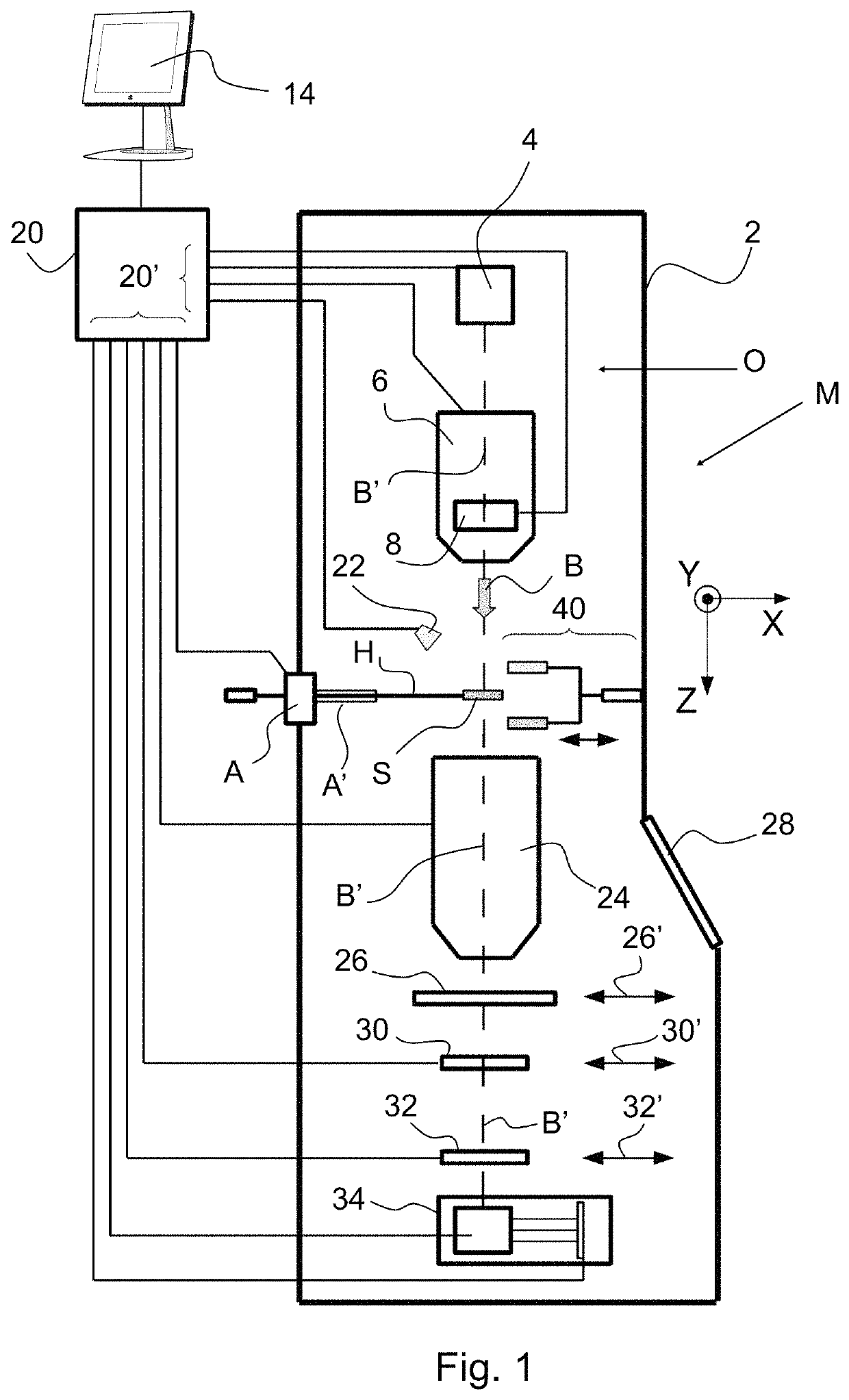
[0038]FIG. 1 (not to scale) is a highly schematic depiction of a charged-particle microscope M, as an example of a possible embodiment of the apparatus that may be used to perform the method as disclosed herein. More specifically, it shows an embodiment of a transmission-type microscope M, which, in this case, is a TEM / STEM (though, in the context of the current invention, it could just as validly be a SEM—see FIG. 2—, or an ion-based microscope, for example). In FIG. 1, within a vacuum enclosure 2, an electron source 4 produces a beam B of electrons that propagates along an electron-optical axis B′ and traverses an electron-optical illuminator 6, serving to direct / focus the electrons onto a chosen part of a specimen S (which may, for example, be—locally—thinned / planarized). Also depicted is a deflector 8, which (inter alia) can be used to effect scanning motion of the beam B.

[0039]The specimen S is held on a specimen holder H that can be positioned in multiple degrees of freedom by...


PUM


Abstract

The invention relates to a screening method. The method comprises the step of providing a sample, wherein said sample comprises a sample carrier with a surface structure, as well as an object of interest. The method further comprises the step of acquiring an image of said sample. According to the disclosure, the method comprises the steps of providing information on said surface structure of said sample carrier, which may in particular comprise the step of acquiring an image of said sample carrier. In that case two images are obtained: one more sensitive to the objects of interest, and one more sensitive to the surface structure of the sample carrier. This allows manipulation of the acquired image, using said information on the surface structure of the sample carrier. With this, said manipulated image may be screened for easy and reliable detection of said object of interest.

Description

BACKGROUND OF THE INVENTION[0001]The present invention relates to a screening method and apparatus for detecting an object of interest. In an embodiment, the present invention is related to the detection of asbestos fibers using a charged particle microscope.[0002]Screening samples for detecting a particular object of interest can be a tedious task. This is in particular the case when a sample is screened for certain objects that are not expected to be there.[0003]For example, in the food and drink industry, samples are gathered from a product, and these products are screened for contaminations, such as, for example, bacteria. In a known prior art method, a sample is obtained from a product, the sample is incubated to allow time for (undesired) bacteria to grow, and then the sample is screened for the presence of these bacteria. When the number of bacteria is below a certain threshold, then this indicates that the product (and the corresponding batch of products) is safe.[0004]In a ...

Claims


Application Information

Patent Timeline
Patent Type & Authority Patents(United States)
IPC IPC(8): H01J37/24H01J37/244
CPCH01J37/244H01J2237/0492H01J2237/2448H01J2237/2449H01J2237/28G01N23/2251G01N2223/079G01N2223/401G01N2223/418G01N2223/42G01N2223/651G06T7/0004G06T7/12G06T2207/10056G06T2207/10116G06T2207/20061G06T2207/20084
Inventor ARTS, WOUTER
Owner PHENOM WORLD HLDG BV
Why Eureka
Try Eureka