
Methods and systems for a single-bolus angiography and perfusion scan

a single-bolus angiography and perfusion scan technology, applied in the field of non-invasive diagnostic imaging, can solve the problem of taking five minutes to achieve the effect of being alon

Active Publication Date: 2021-11-23
GE PRECISION HEALTHCARE LLC
View PDF22 Cites 2 Cited by
  • Summary
  • Abstract
  • Description
  • Claims
  • Application Information

AI Technical Summary

Benefits of technology

This approach ensures diagnostic-quality images by optimizing contrast agent timing, reducing radiation exposure, and expediting patient care in acute stroke management.

Problems solved by technology

However, the timing bolus scan alone takes five minutes, and performing CTA and CTP studies separately requires five to seven minutes between acquisitions to allow contrast washout.

Method used


Image

  • Methods and systems for a single-bolus angiography and perfusion scan
  • Methods and systems for a single-bolus angiography and perfusion scan
  • Methods and systems for a single-bolus angiography and perfusion scan

Examples


Embodiment Construction

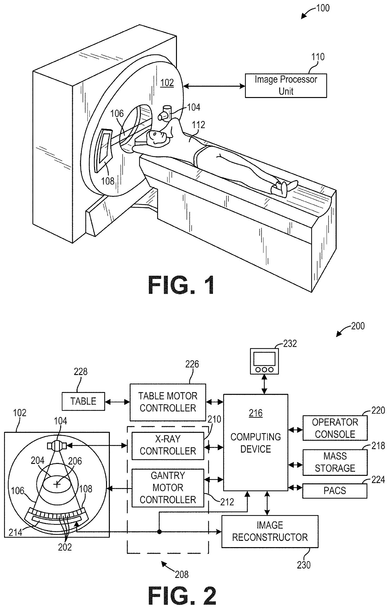
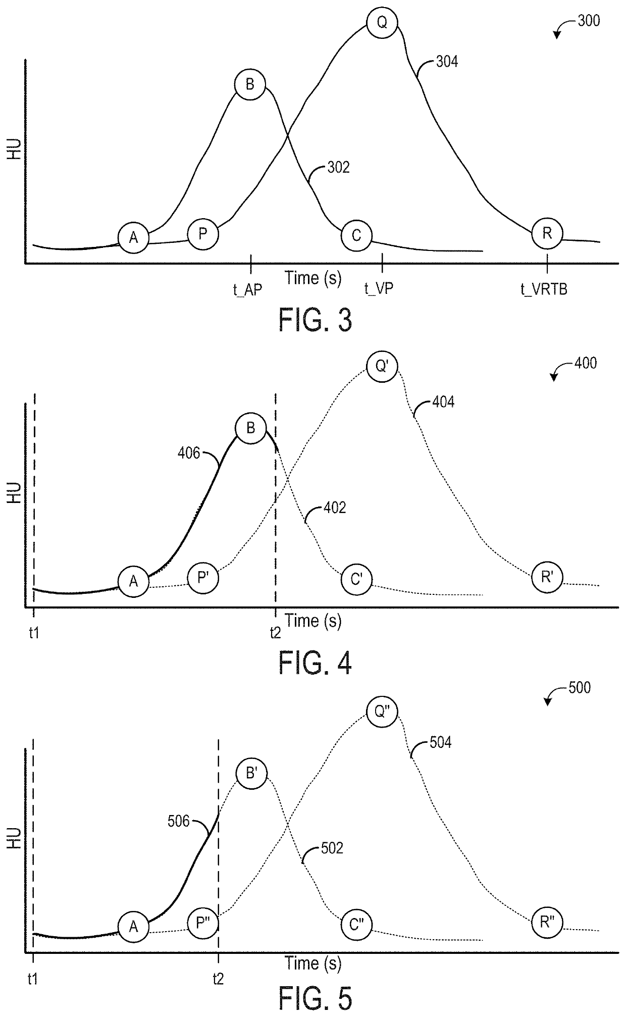
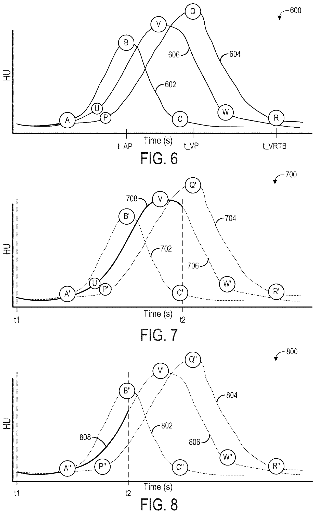
[0027]Some diagnostic imaging protocols, such as protocols to diagnose acute stroke in a patient, include one or more contrast scans, where a contrast agent is administered to the patient prior to the diagnostic imaging scan. These diagnostic imaging protocols may include two contrast scans, such as a computed tomography (CT) angiography (CTA) scan followed by a CT perfusion (CTP) scan. In a CTA followed by a CTP (or in a CTP followed by a CTA), the decision of when to administer the second contrast agent bolus may be challenging, and if timed incorrectly, may result in non-diagnostic images and / or undesired patient outcomes. For example, if the second contrast agent bolus is administered too soon after the first contrast scan, diagnostic image quality of images acquired during the second contrast scan may be degraded due to venous contrast contamination from the first contrast agent bolus. However, if the second contrast agent bolus is administered too late after the first contrast...


PUM


Abstract

Methods and systems are provided for adaptive scan control. In one embodiment, a method includes, upon an injection of a contrast agent, performing a plurality of perfusion acquisitions of a first anatomical region of interest (ROI) of a subject with the imaging system, processing projection data of the first anatomical ROI obtained from the plurality of perfusion acquisitions to measure a contrast signal of the contrast agent, performing a plurality of angiography acquisitions, each angiography acquisition performed at a respective time determined based on the contrast signal, and performing one or more additional perfusion acquisitions between each angiography acquisition.

Description

FIELD[0001]Embodiments of the subject matter disclosed herein relate to non-invasive diagnostic imaging, and more particularly, to real-time adaptive contrast imaging.BACKGROUND[0002]Non-invasive imaging technologies allow images of the internal structures of a patient or object to be obtained without performing an invasive procedure on the patient or object. In particular, technologies such as computed tomography (CT) use various physical principles, such as the differential transmission of x-rays through the target volume, to acquire image data and to construct tomographic images (e.g., three-dimensional representations of the interior of the human body or of other imaged structures).[0003]For emergency room (ER) stroke management, time is critical to determine a proper course of treatment. For every minute a large vessel ischemic stroke is untreated, the average patient loses 1.9 million neurons. For each hour in which a treatment fails, the patient loses as many neurons as it do...

Claims


Application Information

Patent Timeline
Patent Type & Authority Patents(United States)
IPC IPC(8): A61B6/00A61B6/03G06T7/00G06T11/00
CPCA61B6/507A61B6/032A61B6/481A61B6/504G06T7/0012G06T11/003G06T2207/20084G06T2207/30104G06T2211/404A61B6/482G06T11/008G06T2211/424G06T2211/421G06T2211/441
Inventor VAZ, MICHAEL SARJUTALASILA, NITYAGABRIELSE, BRADLEYFORBES, RYANPITTERLE, DAVID JOSEPH
Owner GE PRECISION HEALTHCARE LLC
Learn More