Vision catheter

a catheter and vision technology, applied in the field of medical devices, can solve the problems of large size of the camera, large area, and complex movement type, and achieve the effects of increasing the field of view, and being easy to be assembled

Inactive Publication Date: 2005-02-03
SCI MED LIFE SYST
View PDF6 Cites 110 Cited by
  • Summary
  • Abstract
  • Description
  • Claims
  • Application Information

AI Technical Summary

Benefits of technology

[0010] In accordance with another aspect of the invention, a system that can steer the distal end of the fiber bundle or single fiber is utilized to steer or increase the field of view without moving the device. Whether an imaging lens is utilized on the tip of the bundle, or a fixed objective lens is used on the distal tip of the catheter or guidewire that creates the image plane to be scanned by the fiber bundle, the steering of the distal end of the bundle further increases the field of view.
[0011] It will be appreciated that the vision catheter of the present invention includes components that are widely available and that can easily be assembled. The simple design thus allows for the production of catheters that are relatively inexpensive and disposable and which have imaging capabilities while still remaining relatively small in diameter.

Problems solved by technology

One drawback of this alternative is that such cameras are relatively large in size, in comparison to the dimensions needed for certain surgical procedures.
Another issue with either the semiconductor-type camera or the bundle of fibers, is that the ability to see a larger area requires moving the camera or the bundle of fibers.
This type of movement is relatively complex to implement, and requires even more area.
Furthermore, while endoscopes are a proven technology, they are relatively complex and expensive to manufacture.

Method used


Image

  • Vision catheter
  • Vision catheter
  • Vision catheter

Examples


Embodiment Construction

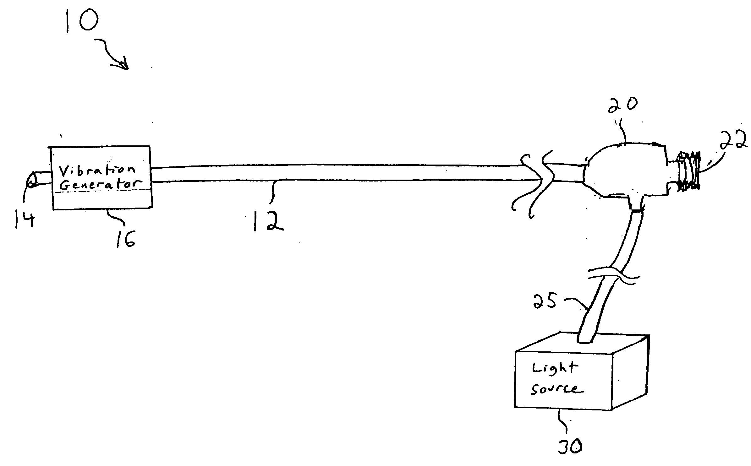
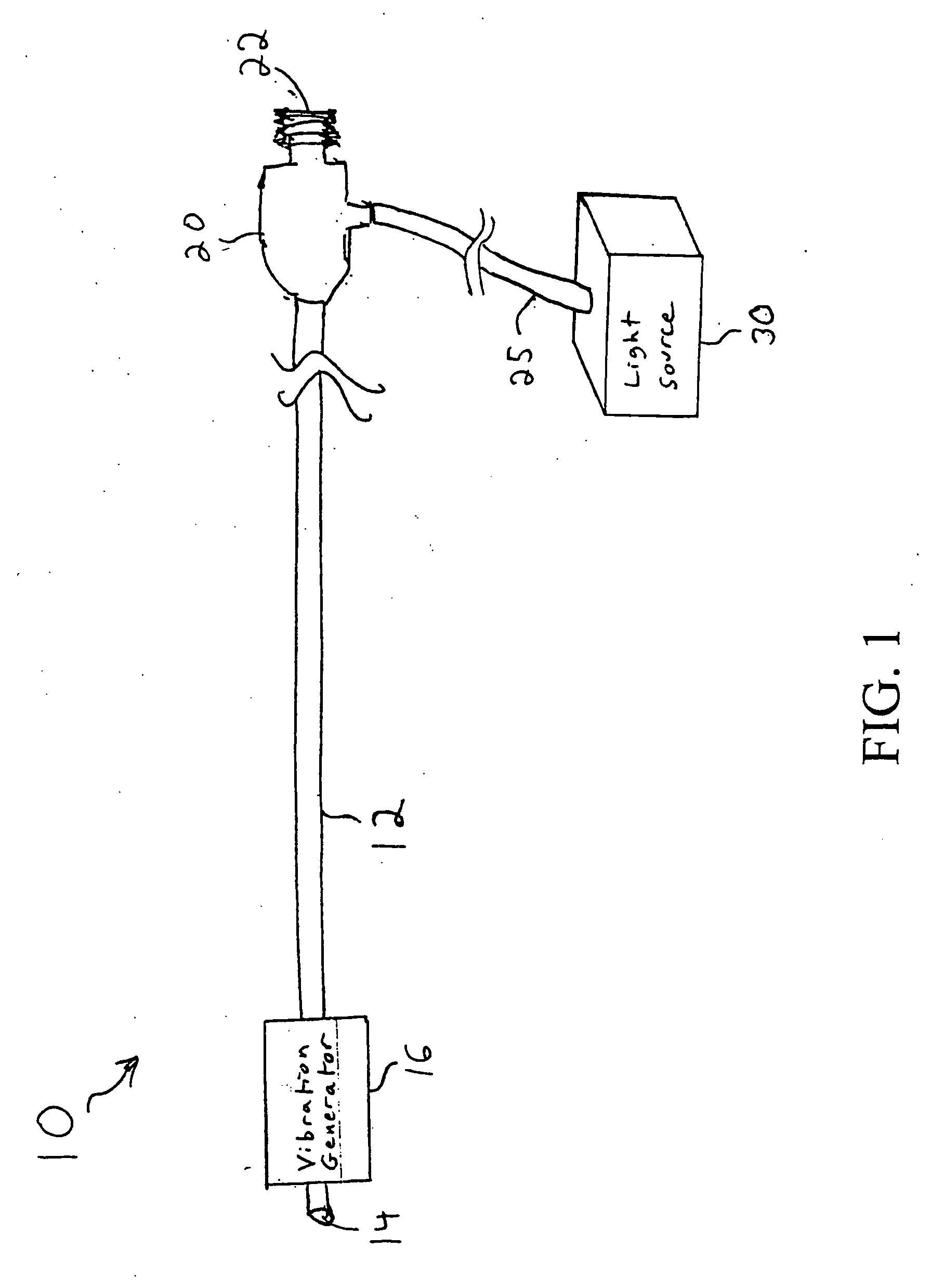
[0018]FIG. 1 is a diagram of a vision catheter 10 formed in accordance with the present invention. The vision catheter 10 includes a flexible imaging cable 12 having a polished distal end 14. In one embodiment, the flexible imaging cable 12 may include a group of standard clad optical fibers. In general, the optical fibers will include one or more imaging fibers and one or more illumination fibers. The imaging fibers transmit image information detected at the distal end 14 of the imaging cable 12. The illumination fibers are coupled to a light source so as to provide illumination at the distal end 14 of the imaging cable 12.

[0019] The vision catheter 10 also includes a vibration generator 16. In accordance with the present invention, the vibration generator 16 vibrates the distal end 14 of the imaging cable 12. This essentially produces a scanning effect in that as the distal end 14 moves, the field of view that is sensed by the distal end 14 effectively increases. As will be descr...


PUM


Abstract

A catheter with a small optical fiber or bundle of fibers includes a scanning mechanism constructed with the use of any vibration capable component. Magnetic, piezoelectric or other mechanisms are used to vibrate the end of the fiber and thus create a scanning effect which extends the field of view. One or more lenses may be utilized, including a lens attached to the distal tip of the image fiber, or a lens attached to the distal tip of the catheter for creating an image plane which can be scanned by the fiber. In one embodiment, multiple light sources may be connected to the fiber for enabling the use of field sequential color techniques for real-time imaging, as well as real-time fluorescent imaging for disease detection. A photodetector assembly connected to the proximal end may contain both filtered and unfiltered detectors for use with both standard imaging and fluorescent imaging. The resulting vision catheter is relatively inexpensive and disposable.

Description

CROSS-REFERENCE TO RELATED APPLICATION [0001] This application is a continuation-in-part of U.S. Patent Application Ser. No. 10 / 630,440, filed Jul. 29, 2003, priority from the filing date of which is hereby claimed under 35 U.S.C. § 120.FIELD OF THE INVENTION [0002] The present invention relates to medical devices, and in particular to a catheter with imaging capabilities. BACKGROUND OF THE INVENTION [0003] An endoscope is a type of catheter that has imaging capabilities so as to be able to provide images of an internal body cavity of a patient. Most minimally invasive surgical procedures performed in the GI tract or other internal body cavities are accomplished with the aid of an endoscope. A typical endoscope has an illumination channel and an imaging channel, both of which may be made of a bundle of optical fibers. The illumination channel is coupled to a light source to illuminate an internal body cavity of a patient, and the imaging channel transmits an image created by a lens ...

Claims


Application Information

Patent Timeline
Patent Type & Authority Applications(United States)
IPC IPC(8): A61B1/005A61B1/04A61B1/07
CPCA61B1/0008A61B1/00096A61B1/00165A61B1/07A61B1/00183A61B1/0051A61B1/00172
Inventor BARBATO, LOUIS J.HAMM, MARK A.CHIN, YEM
Owner SCI MED LIFE SYST
Why Eureka
Try Eureka