
Method and arrangement for reducing the volume of a lung

a technology of lung volume and arrangement, applied in the direction of respirator, respirator, fire rescue, etc., can solve the problem of short pressure peak

Inactive Publication Date: 2005-03-24
PULMONX
View PDF12 Cites 200 Cited by
  • Summary
  • Abstract
  • Description
  • Claims
  • Application Information

AI Technical Summary

Benefits of technology

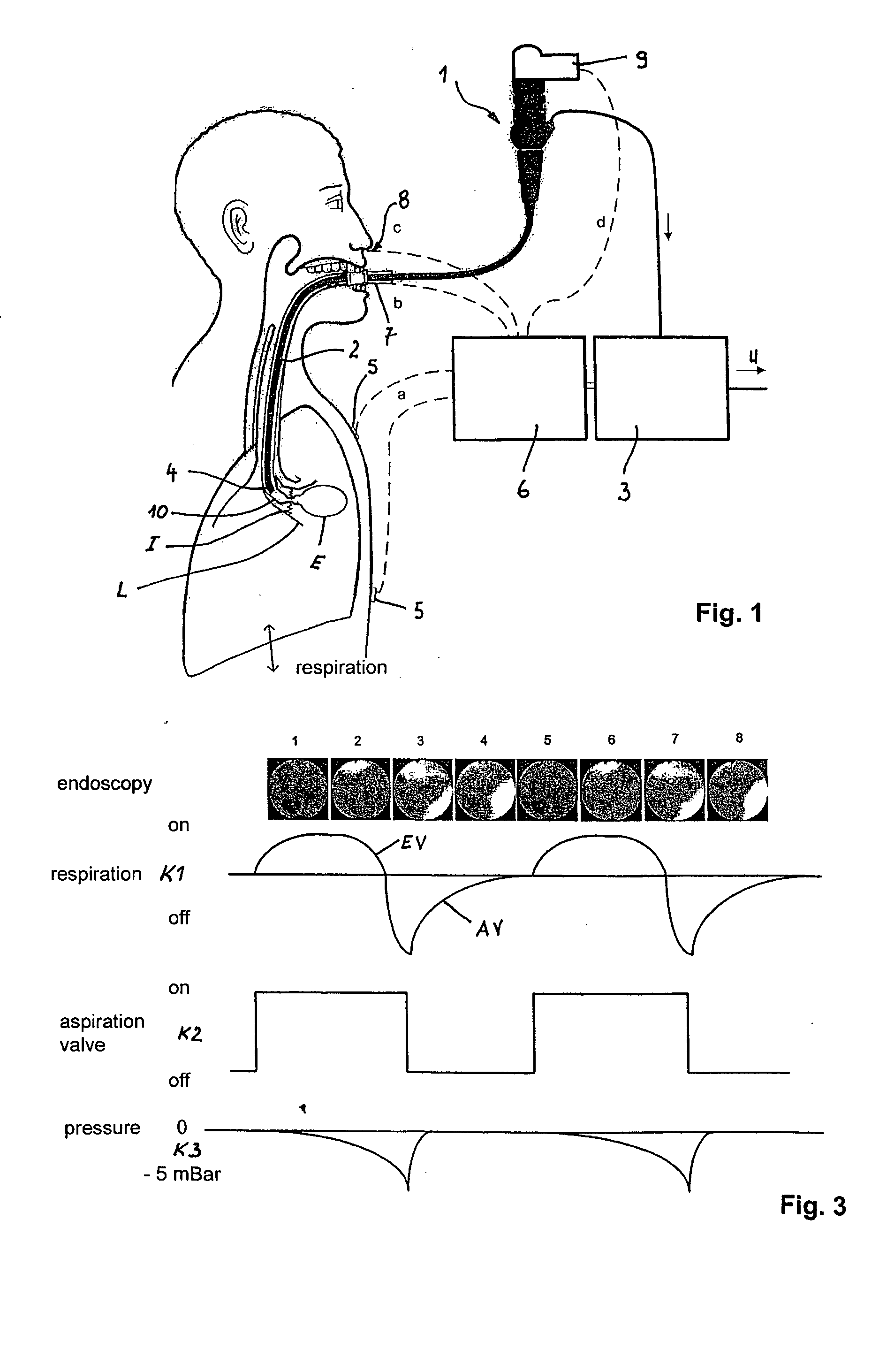
[0009] Methods and apparatus according to the present invention inhibit collapse of the segmental bronchus or lung tissue during aspiration associated with lung volume reduction procedures. More specifically, the methods and apparatus of the present invention provide for aspiration synchronous with the patient's respiration cycle to remove air during periods of patient inhalation when the bronchus or airways leading to or within the hyperextended lung region being treated are generally open and available to transport air from the region. Conversely, aspiration is not performed during patient exhalation when the airways leading to and / or within the hyperextended lung region may be subject to collapse which would prevent or inhibit air transport from the region. Alternatively or additionally, airway collapse can be inhibited or reversed by short pulses of pressurized gas.
[0010] A bronchial catheter is introduced into a hyperexpanded lung area, and air is aspirated from there by means of an aspiration device. During treatment, the patient's spontaneous respiration is recorded. This can be done manually, but preferably is accomplished automatically using sensors and measuring devices. Aspiration of the air from the emphysematous or otherwise hyperextended lung region is carried out in synchrony with the patient's inhalation action. The invention thus makes use of the characteristic that that the lung is expanded during inhalation. The lung draws the bronchi away from one another. This phenomenon is known as interdependence. According to the invention, it is in this expanded state during inspiration or inhalation that aspiration of the isolated region to be treated is carried out. In this way, the risk of collapse of the surrounding airways upon application of an underpressure can be lessened.
[0011] In an alternative aspect of the present invention, the bronchus leading to or within the isolated region to be aspirated may be widened by a compressed gas pulse during aspiration of the air. By pulsing compressed gas, the airways adjacent to the distal end of the bronchial catheter are widened and opened prior to or during the aspiration procedure. Optionally, potential collapse of the bronchus or airways may be visually or otherwise monitored, and a short overpressure pulse expediently delivered whenever a potential collapse is detected. The action of the compressed gas results in short pressure peaks. By this means, the bronchus can be widened exactly at the time of a collapse. This allows the desired aspiration to be carried out.
[0017] Preferably the aspiration procedure should not be carried out when the affected segmental bronchus collapses, and, in the event of a collapse, the volume should be expanded by means of a compressed gas stream. To determine the actual situation in the body during treatment, an image can also be recorded in situ. An imaging unit may form a component part of the system and be linked to a data processing unit for controlling the pressure generator. The images are continuously monitored, and the image information is then converted to digital signals and, if appropriate after contrast enhancement, used to evaluate the state in the lung area. In this way, a collapse, or an imminent collapse, can be detected, and a suitable compressed gas pulse can be generated in good time.

Problems solved by technology

Conversely, aspiration is not performed during patient exhalation when the airways leading to and / or within the hyperextended lung region may be subject to collapse which would prevent or inhibit air transport from the region.
The action of the compressed gas results in short pressure peaks.

Method used


Image

  • Method and arrangement for reducing the volume of a lung
  • Method and arrangement for reducing the volume of a lung
  • Method and arrangement for reducing the volume of a lung

Examples


Embodiment Construction

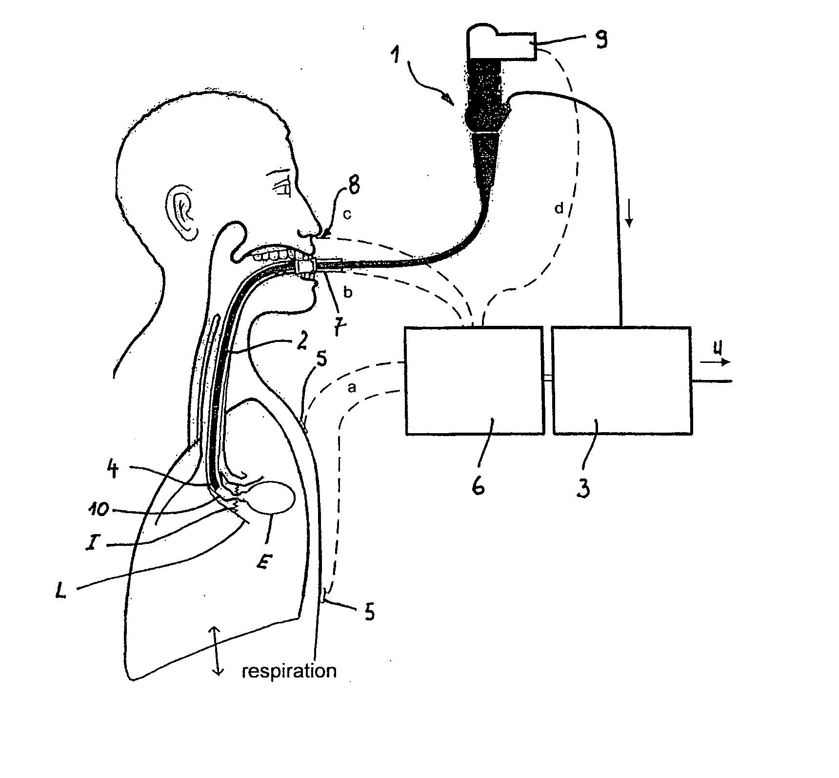
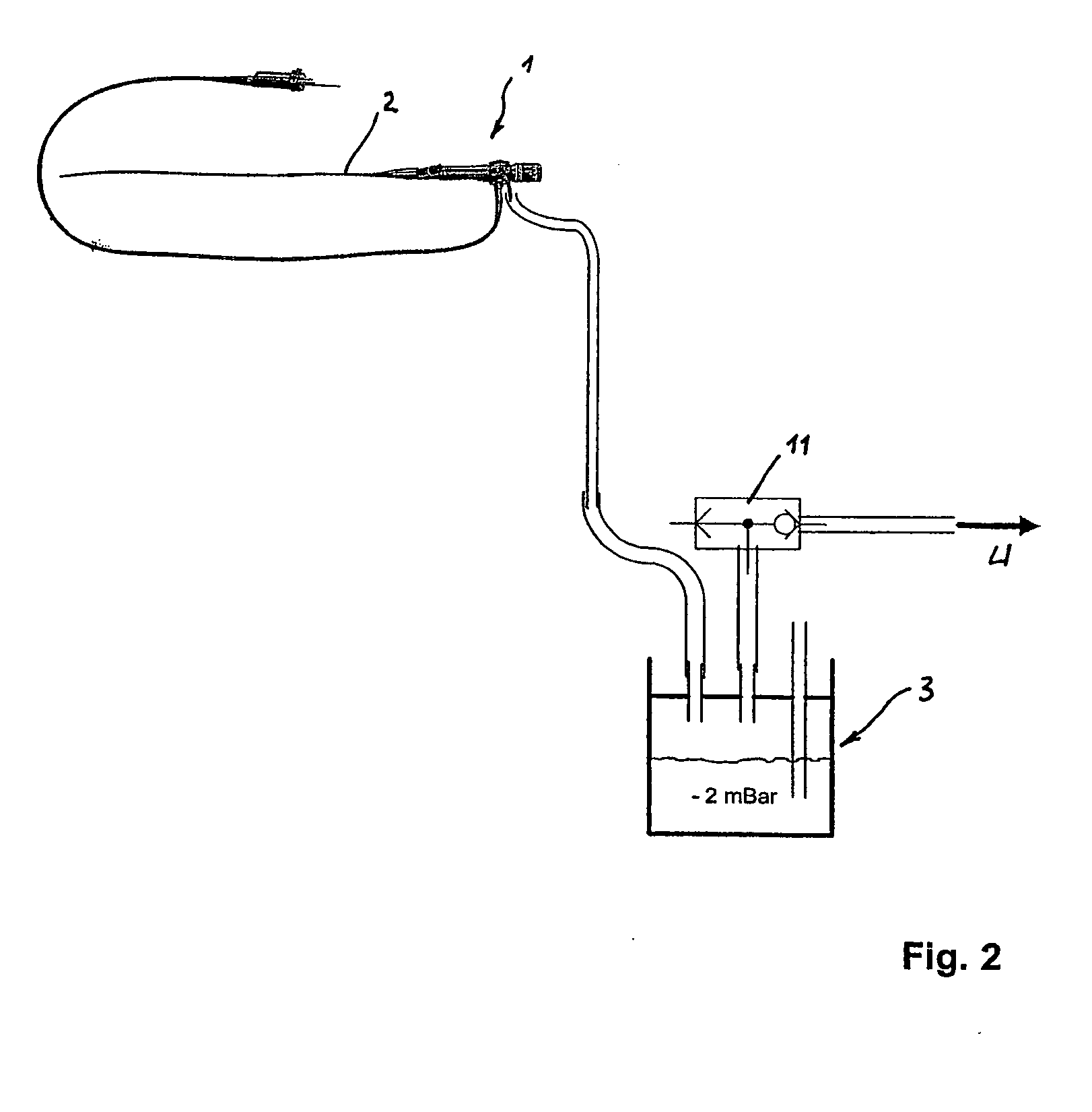
[0027]FIG. 1 shows a diagrammatic representation of an arrangement according to the invention for reducing the volume of a lung L of a patient, who suffers from pulmonary emphysema, during treatment. The lung area affected by the emphysema is designated by E. The basic structure of the arrangement can be seen from FIG. 2.

[0028] The arrangement comprises a bronchoscope 1 with a bronchial catheter 2 which communicates with an aspiration device 3. The bronchial catheter 2 is introduced into the hyperexpanded lung area. There, the distal end 4 of the bronchial catheter 2 can be sealed off relative to the surrounding vessel wall by means of suitable blockers (not shown here). Sensors 5 secured on the patient's chest record the patient's spontaneous respiration by measuring the thorax impedance. The measurement values recorded by the sensors 5 are evaluated by computer in a control unit 6, forming a component part of the aspiration device, 3 and are used for controlling the aspiration pr...


PUM


Abstract

The invention relates to a method and an arrangement for reducing the volume of a patient's lung. A bronchial catheter (2) is introduced into a hyperexpanded lung area, and air is aspirated from there by means of an aspiration device (3). The associated segmental bronchus is then closed. According to the invention, the patient's spontaneous respiration is recorded by sensors (5), and aspiration of the air is carried out in synchrony with the patient's inhalation action. In order to prevent collapse of the associated segmental bronchus, a pressure generator is provided with which the associated segmental bronchus can be widened, by a compressed gas pulse, in synchrony with the aspiration. The pressure generator can be activated as a function of the aspirated air stream, which is monitored with a measuring device.

Description

CROSS-REFERENCES TO RELATED APPLICATIONS [0001] This application is a continuation-in-part of PCT / DE2004 / 000008 (Attorney Docket No. 017354-002700PC), filed on Jan. 7, 2004, which claimed priority from DE10302310.0, filed on Jan. 20, 2003, the full disclosures of which are incorporated herein by reference.BACKGROUND OF THE INVENTION [0002] Field of the Invention [0003] The invention relates to a method and an arrangement for reducing the volume of a lung in a patient suffering from pulmonary emphysema. [0004] Pulmonary emphysema is, in general terms, a hyperexpansion of the lung tissue. It develops when pulmonary alveoli and terminal bronchioles burst and are destroyed, so that, instead of a large number of small pulmonary alveoli, a small number of large air cells, or regular sacs, develop. This leads to a reduction in the surface area for gas exchange. This means that the capacity for intake of oxygen and release of carbon dioxide is then much lower. Even very slight physical exer...

Claims


Application Information

Patent Timeline
IPC IPC(8): A61M16/04
CPCA61M16/0404A61M16/04
Inventor FREITAG, LUTZ
Owner PULMONX
Learn More